Cores Directory
Search for the resources you need to answer your research questions.
Active Filters:
Listing of research core items
-
Instrument/equipment
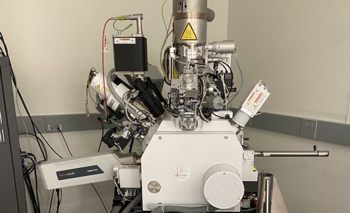
Helios G4 UX DualBeam System
Helios G4 UX DualBeam (electron & ion-beam) microscope
Located within Aberration-Corrected Electron Microscopy (ACEM) Core -
Instrument/equipment
Imaris image analysis workstations
Imaris imaging analysis workstations
Located within Confocal Microscopy at CCMI -
Core/facility
In Vivo Imaging (IVI) Facility
The core offers researchers access to an upright two-photon laser scanning microscope optimized for in vivo fluorescence imaging studies. Training or direct staff assistance is available in surgical preparations, imaging acquisition, and data analysis.
David Gonzalez, MHS LAT
Facility Manager -
Instrument/equipment
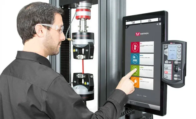
Instron tensile/compression testing
This instrument performs tensile, compression, flexure/bend, peel, tear, shear, and cyclic tests.
Located within Mechanical and Thermal Analysis Instrumentation Core (MTAIC) -
Services
Instrument design & electronics prototyping
We have designed instruments and parts for groups across campus and across disciplines. We also have some capacity for prototyping and development of electronics for instrumentation work.
Located within Advanced Prototyping Center at Wright Lab -
Instrument/equipment
Ion mill
The Hitachi IM4000 can be used for cross-sectioning samples (usually for SEM viewing) or for polishing surfaces.
-
Instrument/equipment
Iridium sputtering tool
Iridium is useful for coating samples before electron microscopy.
-
Services
Isotope ratio mass spectrometry (IRMS)
Isotope ratio mass spectrometry (IRMS) services
Located within Yale Analytical and Stable Isotope Center (YASIC) -
Instrument/equipment
Laser cutting
Our medium-power laser cutter can be best thought of as a computer-controlled bandsaw. Like the abrasive water jet, it is largely used to cut through flat material stock. It can also be used to etch various materials.
Located within Advanced Prototyping Center at Wright LabCraig Miller
-
Instrument/equipment
Leica SP5 confocal microscope
Leica SP5 confocal microscope
Located within Confocal Microscopy at CCMIMateus Guerra, PhD
Primary contact Associate Director -
Instrument/equipment
Leica SP8 Gated STED 3X super resolution
Leica SP8 Gated STED 3X Super Resolution microscope
Located within Confocal Microscopy at CCMIMateus Guerra, PhD
Primary contact Associate Director -
Instrument/equipment
Leica STELLARIS 8 TauSTED confocal microscope
Leica DM6 CS upright microscope with environmental control system.
Located within Light Microscope Imaging Facility on Science HillJoseph Wolenski, PhD