In Vivo Imaging (IVI) Facility
David Gonzalez, MHS LAT
Facility ManagerBuilding: TAC, rooms S-614 and S-600
About the core
The core provides support for Yale investigators interested in utilizing two-photon fluorescence microscopy to achieve their research goals. It is responsible for novel protocol and methods development and is designed to serve as a clearinghouse for expertise and experience, being able to translate newly successful approaches to multiple projects and investigators.
It is the core’s goal to assist investigators in experimental design and implementation and to provide training for those whom wish to learn surgical preparations, imaging acquisition, and data analysis at the facilities image workstation utilizing Imaris software (Oxford Instruments).
- Microscope suite
- LaVision Biotec TriMScope
- PC workstation with Imaris software (Oxford Instruments)
Services include specimen preparation, user training, and advice on experimental design.
Available to Yale researchers only
Core websiteContacts
-
Core/facility
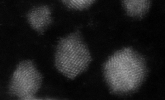
Aberration-Corrected Electron Microscopy (ACEM) Core
Our core's focus is cutting-edge and high-throughput capability in electron microscopy techniques, including TEM, SEM, and FIB.
-
Core/facility
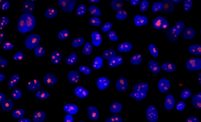
Yale Center for Molecular Discovery
We provide resources for assay development and high-throughput screening with small molecules and arrayed siRNA and sgRNA/CRISPR libraries in biochemical, cell-based and image-based assays (primarily 384-well format).
-
Core/facility
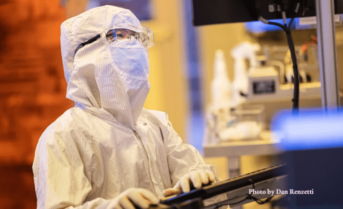
Yale Cleanroom Facilities
The University Cleanroom provides a controlled environment that maintains intentionally low concentrations of pollutants to allow for uncontaminated scientific research.
Yong Sun & Lauren McCabe