Cores Directory
Search for the resources you need to answer your research questions.
Active Filters:
Listing of research core items
-
Core/facility
Earth Materials Characterization Center (EMC2)
We offer materials physics equipment including microscopy, spectroscopy, deformation tools, and sample-prep tools for sample characterization work.
-
Instrument/equipment
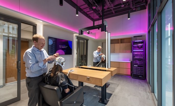
Electroencephalography (EEG)
Electroencephalography (EEG) reveals brain function with millisecond precision by measuring, at the scalp, the minute magnetic fields generated by neural activity.
Located within BrainWorks -
Core/facility
Electron Microscopy Facility at CCMI
The Electron Microscopy Facility at the Center for Cellular and Molecular Imaging (CCMI) provides investigators with a wide range of electron microscopy services.
-
Instrument/equipment
EM CCMI instruments
Microscopes, sample preparation, carbon evaporators, and advanced cryo FIB lamella milling for cryo electron tomography.
Located within Electron Microscopy Facility at CCMI -
Services
EM CCMI services
Project consultation and experimental design, sample preparation, and imaging with transmission and scanning electron microscopes.
Located within Electron Microscopy Facility at CCMI -
Instrument/equipment
Eye tracking
Our EyeLink 1000 Plus systems offers high-speed, precise eye tracking with flexible head-fixed and head-free modes for cognitive and vision research.
Located within BrainWorks -
Core/facility
FIB-SEM Collaboration Core (F-SCC)
We utilize unparalleled enhanced FIB-SEM pipeline to generate large-volume three-dimensional electron microscopy data with nanometer isotropic resolution. Such data reveal critical insights about structure-function relationships in biology.
-
Instrument/equipment
Functional near-infrared spectroscopy (fNIRS)
Functional near-infrared spectroscopy (fNIRS) is a portable technique that uses near-infrared light to estimate cortical hemodynamic activity related to neural activity.
Located within BrainWorks -
Instrument/equipment
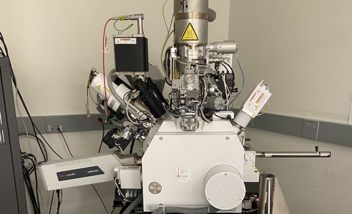
Helios G4 UX DualBeam System
Helios G4 UX DualBeam (electron & ion-beam) microscope
Located within Aberration-Corrected Electron Microscopy (ACEM) Core -
Instrument/equipment
Imaris image analysis workstations
Imaris imaging analysis workstations
Located within Confocal Microscopy at CCMI -
Core/facility
In Vivo Imaging (IVI) Facility
The core offers researchers access to an upright two-photon laser scanning microscope optimized for in vivo fluorescence imaging studies. Training or direct staff assistance is available in surgical preparations, imaging acquisition, and data analysis.
David Gonzalez, MHS LAT
Facility Manager -
Instrument/equipment
Ion mill
The Hitachi IM4000 can be used for cross-sectioning samples (usually for SEM viewing) or for polishing surfaces.