FIB-SEM Collaboration Core (F-SCC)
Building: SHM B-wing, room E10
About the core
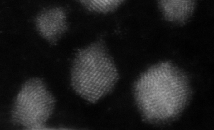
At the forefront of volume electron microscopy (EM), we welcome your collaboration to take discoveries in biology, physiology, and pathology to a whole new level using our proprietary enhanced FIB-SEM (eFIB-SEM) technology and its unparalleled pipeline. This enhanced FIB-SEM technology enables 3D large-volume high-resolution imaging, capable of years of continuous imaging without defects in the final image stack.
Offerings
- EM sample preparation with TEM evaluation
- Non-destructive 3D X-ray tomography
- X-ray guided eFIB-SEM sample preparation to define, reposition, and refine region of interest (ROI)
- 3D large-volume isotropic high-resolution eFIB-SEM imaging
- eFIB-SEM image registration and alignment
- eFIB-SEM data segmentation and analysis (to be announced)
Available to Yale researchers & external researchers
Core websiteContacts
Physical address:
SHMB E10
333 Cedar Street
New Haven, CT 06510
Postal address (USPS):
P.O. Box 208026
333 Cedar Street
New Haven CT 06520-8026
Shipping address (FedEx and UPS):
c/o Cellular & Molecular Physiology Department
SHMB E36A
200 South Frontage Rd
New Haven, CT 06510

