Electron Microscopy Facility at CCMI
Building: SHM, suites IE-26, 38, & 40
About the core
The Center for Cellular and Molecular Imaging (CCMI) Electron Microscopy Facility is a central core facility administrated and financially supported by the Dean’s office at Yale Medical School. We provide expertise, instrumentation, and training in modern electron microscopy, and work closely with investigators on their science projects. The facility is located at Sterling Hall of Medicine (333 Cedar Street), suite IE-26, 38 and 40.
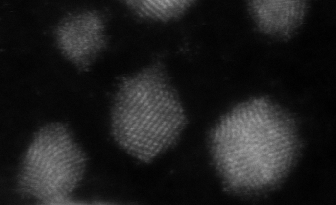
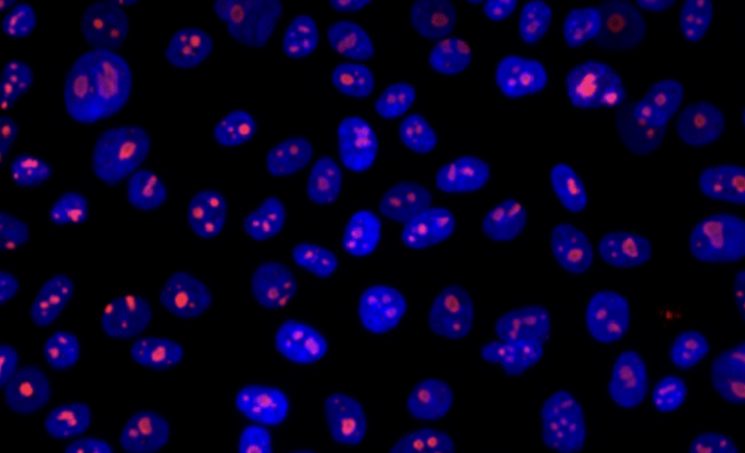
The core provides a wide range of electron microscopy services. These include sample preparation, standard transmission and scanning electron microscopy for ultrastructure, high-resolution protein localization, tomographic 3D rendering of the whole cell using focused-ion-beam scanning electron microscopy (FIB-SEM), cryo FIB milling, lamellae prep, and correlative light and electron microscopy (CLEM), which are growing in demand. We work closely with investigators and also train them to use the facility's instruments independently.
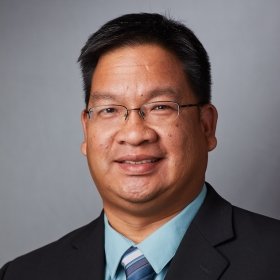
Anyone who wishes to start an EM project or to access facility equipment should contact the facility director Xinran Liu at 203-785-4050 or by email.
Available to Yale researchers & external researchers
Starting an EM project
For further details on our full-service EM support, use of facility equipment, and training, please contact the facility director, Xinran Liu, to schedule a consultation meeting.
Policies
The instruments in the facility represent a substantial investment and resource for the research community. To ensure that these resources are available to investigators at the optimum performance level and a minimum of down-time, the following policies have been established.
In order to gain access to the EM core facility and its instruments, investigators must meet with the director of the facility and, in case training is required, be trained by the staff.
If you experience any problems while operating instruments, please record them in the log book and notify the facility staff immediately.
Instruments and chemicals cannot be removed from the lab at any time. Liquid nitrogen in the lab should only be used for electron microscopy applications. Computers in the microscope rooms are solely used for imaging purposes. The computers that operate the microscopes should never be used to browse the internet and write/read emails.
Acquired digital images are stored on the facility image server and can be accessed through the Yale network. However, due to limited hard drive space, this storage is meant to be temporary. Old files will be deleted periodically without warning.
Use of negative staining is confined to the specific bench space. Hazardous and radioactive waste must be properly disposed of in the assigned containers. Due to limited space, users should not store their samples, solutions, or other materials (grids, blocks, etc.) in the facility unless authorized by staff.
Users who abuse policies and/or lack respect towards other users or the facility staff may result in access to the facility being revoked.
Microscopy and instrument reservation
Microscopes are made available on a first-come, first-served basis. Contact staff to reserve instruments or use the online scheduling system.
Use of the facility by fully trained individuals is allowed after hours and on weekends. In case of an emergency, the telephone numbers of the director and other personnel are posted at various locations around the lab. Access during regular hours is preferred due to the available support.
Safety
The staff will provide instructions on how to handle chemicals and instruments in a safe manner. Users who will be exposed to uranyl acetate solutions need to register for a class in radiation safety before being allowed to start their work in the lab. No other radioactive materials are allowed in the EM facility. Users need to inform the staff before attempting to submit samples that contain radioisotopes.
All samples that are submitted to the EM facility need to be previously fixed with either paraformaldehyde or glutaraldehyde, so as to ensure that any pathogens they might contain have been inactivated.
Spills of chemicals need to be notified and cleaned immediately. In addition, users should take great care in cleaning the bench space and instruments right after use.
Cancellation & tardiness
Cancellation: If for any reason you cannot use your session, it is your responsibility to delete the sign-up entry from the scheduler as early as possible and notify the facility staff.
Tardiness: If a user does not show up within one hour of the starting time, the reservation will be lost. The slot is then open to other users to sign up. Tardiness without notice will be fully billed for the entire period.
Acknowledgments
Proper acknowledgment enables us to obtain financial and other support, so we can continue to provide the cutting-edge technologies and instruments for your research. If you are publishing or presenting data acquired in the core facility, please acknowledge our contribution. Here is an example: "The authors would like to thank the Center for Cellular and Molecular Imaging, Electron Microscopy Facility at Yale Medical School for assistance with the work presented here.”
Rates
Effective July 1, 2024. See also training rates.
Regular services
Regular services are invoiced monthly after a project is completed.
| Regular services | Yale | Other nonprofits |
|---|---|---|
| TEM sample processing & embedding (1-4) | $377.65 | $510.00 |
| TEM sample processing & embedding (5+) | $105.00 | $150.00 |
| High-pressure freezing (per shot, unassisted) | $32.00 | $50.00 |
| Freeze substitution (per run, unassisted) | $355.00 | $495.00 |
| SEM sample preparation (1-4) | $410.00 | $582.42 |
| FIB-SEM sample preparation (per run) | $675.00 | $1,000.00 |
| Cryo sample processing (each) | $139.00 | $198.51 |
| Cryo ultramicrotome sectioning (per run) | $175.00 | $250.00 |
| Immunogold labeling (per run) | $343.34 | $410.00 |
| Negative staining (per run) | $100.00 | $150.00 |
| User post-imaging assistance (per hour) | $110.00 | $150.00 |
| CLEM workflow (Live-cell to TEM) | $1,000.00 | $1,350.00 |
| Staff assistance (per hour) | $90.00 | $120.00 |
Instrument rates
| Regular services | Yale | Other nonprofits |
|---|---|---|
| FEI Tecnai Biotwin TEM (per hour, unassisted) | $80.00 | $100.00 |
| FEI Tecnai TF20 TEM (per hour, unassisted) | $110.00 | $135.00 |
| Zeiss Crossbeam SEM imaging (per run) | $135.00 | $155.00 |
| Zeiss Crossbeam FIB-SEM data collection (1 day) | $950.00 | $1,025.00 |
| Zeiss Crossbeam FIB-SEM data collection (2 days) | $1,350.00 | $1,850.00 |
| Leica EM ACE200 carbon coating (per run) | $55.00 | $85.00 |
| Lecia UC7 Ultramicrotome (per hour) | $65.00 | $125.00 |
| Thermo Fisher Glacios (per hour, peak/off-peak) | $70.00/$54.00 | $158.00 |
| FEI Tecnai T12 TEM (per hour, peak/off-peak) | $54.00/$43.00 | $121.00 |
| FEI Vitrobot (per hour) | $48.00 | $142.00 |
| Chameleon (per hour) | $80.00 | $200.00 |
| Leica EM ACE600 carbon coating (per hour) | $32.00 | $152.00 |
Training rates
See training ratesContacts

Faculty Director
