West Campus Imaging Core
Building: Integrated Science and Technology Center basement, rms 006 & 008; office, rm 013
About the core
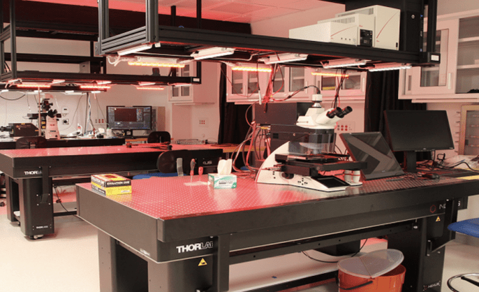
The Imaging Core provides access and training to a broad collection of shared instruments. These include a fleet of optical microscopes with three confocal systems, two wide-field systems, a laser-microdissection system, a cell-culture microscope, a stereomicroscope, and a newly built optical tweezer system. An atomic force microscope (AFM) is also available.
The core also maintains cell-culture equipment, such as a biosafety cabinet, incubators, a refrigerator, and microfuges and lab support areas for cell culture and sample preparation.
Available to Yale researchers & external researchers
PPMS
All users must have an account in our lab management system, PPMS. Internal users can follow the PPMS Quick-Start Manual and External users can use the External User Profile Creation by User document to set up your respective accounts.
Acknowledgment
Please acknowledge the use of all Yale West Campus Imaging Core instrumentation in your research publications and grant applications as follows:
Acknowledgment: This research made use of the Yale West Campus Imaging Core (RRID:SCR_023380).
A copy of the publication should be sent to Joerg Nikolaus for record-keeping purposes. This information is important in helping support our mission and to demonstrate the need for equipment and staff expertise.
Proper reporting of imaging experiments
As microscopy techniques are becoming more powerful and more complex, please also make sure that your publication in a scientific journal contains the full detail about how each image was acquired. Please see the publication by Marques et al. for guidance.
Rates
New instrument rates as of July 1, 2025
| Microscope | Internal Yale ($/hr) |
|---|---|
| MINFLUX -- Abberior | 60 |
| STEDYCON/spinning disk confocal -- Abberior/Nikon | 44 |
| Laser scanning confocal -- Leica Stellaris 8 | 44 |
| Laser scanning confocal -- Leica SP8 | 36 |
| Andor spinning disk confocal -- BC43 | 39 |
| Blaze light sheet -- Miltenyi | 45 |
| Laser microdissection system -- Leica LMD7000 | 27 |
| Widefield -- Leica Thunder Imager 3D Tissue | 26 |
| Widefield -- Leica DMi8 | 21 |
| Optical tweezers | 27 |
| Atomic force -- Asylum Research Cypher ES | 22 |
| IMARIS image analysis workstation | 6 |
Free training for all users.
The Leica DMi8, Leica Thunder, and Leica LMD are charged per actual time used. The Abberior MINFLUX, Abberior/Nikon STEDYCON/SDC, Leica Stellaris 8, Leica SP8, Andor BC43, Miltenyi Blaze, AFM and the Imaris workstation are charged as "earlier of reserved or actual start" until "later of reserved or actual end." MINFLUX users must enlist an advanced imaging scientist of the WCIC to perform experiments on the instrument. Billing is no longer capped.
Other equipment in imaging core (use free of charge)
- Inverted fluorescence microscope Leica DMIL LED
- Stereo microscope Leica M125
Available software on imaging core workstations (room 006)
- Leica LAS X software
- Nikon Elements software and Free viewer
- Optical design software: Zemax
- Mechanical design software: Solidworks
Advisory committee
Andre Levchenko (Systems Biology; Chair)
Julian Berro (Nanobiology)
Erdem Karatekin (Nanobiology)
Ken Loh (Biomolecular Design and Discovery)
John MacMicking (Systems Biology)
Kirstin Meyer (Systems Biology)
Sathish Ramakrishnan (Nanobiology)
Sarah Slavoff (Biomolecular Design and Discovery)
Contacts
Physical address
Yale West Campus Imaging Core
850 West Campus Drive
West Haven, CT 06516
Shipping address
Yale West Campus Imaging Core
WC ISTC, Rm 013
750 West Campus Drive
West Haven, CT 06516

Faculty Director

-
Core/facility
Aberration-Corrected Electron Microscopy (ACEM) Core
Our core's focus is cutting-edge and high-throughput capability in electron microscopy techniques, including TEM, SEM, and FIB.
-
Core/facility
Yale Center for Molecular Discovery
We provide resources for assay development and high-throughput screening with small molecules and arrayed siRNA and sgRNA/CRISPR libraries in biochemical, cell-based and image-based assays (primarily 384-well format).
-
Core/facility
Yale Cleanroom Facilities
The University Cleanroom provides a controlled environment that maintains intentionally low concentrations of pollutants to allow for uncontaminated scientific research.
Yong Sun & Lauren McCabe