Super-resolution and confocal microscopes
Building: Integrated Science and Technology Center basement, rooms 006 & 008; office, room 013
About the instruments
Abberior STEDYCON
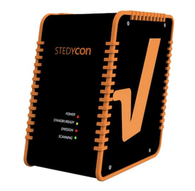
From the manufacturer: "The STEDYCON upgrades your existing widefield system to a confocal microscope and STED nanoscope with a resolution down to 30 nm. All that’s required is a free camera port and a good objective lens. With its super-intuitive user interface, the STEDYCON provides an intelligent microscope platform that enables everyone to acquire superb superresolution images after only minutes of training.
The incredibly compact STEDYCON has the size of a shoebox, yet it is a full-fledged confocal and STED microscope. It fits on every microscope frame with a camera port.
The STEDYCON is service and maintenance-free: The laser beams are aligned by design. So, there is no need to align the system, ever, and it works reliably every day.
Save your time with the STEDYCON software: only minutes of training are required to acquire STED and confocal images. Thanks to the powerful dye database behind, only minimal user input is needed, and even with unknown samples, you get with 3 clicks to the first STED image!
Our software is browser-based, so run it from any device, from any platform, from anywhere." Learn more.
Andor BC43
From the manufacturer: "This plug-and-play laser confocal system has been designed with cost, performance and accessibility in mind, ensuring hassle-free, high-quality 2D & 3D imaging for busy researchers. BC43 is a super-compact unit that is rich in features and benefits, making it the ideal microscope for early-stage researchers and experienced microscopists alike. With no requirement for a darkroom, researchers can access laser confocal microscopy technology on the same bench as their samples. This, coupled with the fast learning curve, allows individual users and entire labs to become much more productive, without compromising results." Learn more.
Leica SP8
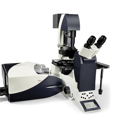
From the manufacturer: "The SP8 LIGHTNING confocal microscope allows you to make proper and detailed observations of fast biological processes. Your experimental work will have the benefit of super-resolution, high-speed imaging, and the capability to image multiple fluorescent markers simultaneously.
Thanks to the exclusive detection concept of LIGHTNING, you can trace the dynamics of multiple molecules, even those expressed at low levels, simultaneously over long recording times in living specimens.
The SP8 LIGHTNING confocal microscope offers you the following performance advantages
- truly simultaneous multicolor imaging in super-resolution down to 120 nm
- live specimen imaging thanks to fast acquisition rates
- sample protection thanks to low phototoxicity
The SP8 LIGHTNING confocal microscope opens up unique experimental options to inspire your research!" See the manufacturer page.
Leica Stellaris 8 Falcon
From the manufacturer: "STELLARIS FALCON (FAst Lifetime CONtrast) is the future of functional imaging. Harness the power of fluorescence lifetime to investigate cellular physiology and explore dynamics in living cells. STELLARIS FALCON is a fully integrated solution for Fluorescence Lifetime Imaging (FLIM) and enables video-rate lifetime imaging acquisition for rapid kinetic studies in live cells.
STELLARIS FALCON adds a new dimension of contrast to your imaging, opening the door to biosensing and tracking of interactions between proteins. FLIM information is now available for all modalities of the STELLARIS system.
Now you can:
- Follow fast molecular interactions via FLIM-FRET (Förster Resonance Energy Transfer)
- Use biosensors to detect changes in metabolic state and microenvironment
- Apply lifetime contrast to separate multiple fluorophores
Acquire FLIM data with minimal training"
Nikon CSU-W1
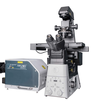
From the manufacturer: "Features a wider field of view and higher image quality than previous models.
- Enhanced signal‐to‐noise ratio
- Ultra‐wide field of view
- Flexible configurations
A new pinhole design significantly reduces pinhole crosstalk, which is particularly important with thicker specimens. The field of view of the CSU-W1 is nearly 4 times that of the CSU-X1, making it ideal for large-area scanning applications and low magnification imaging of large specimens. This large field of view is a perfect match for large format sensors. The CSU-W1 can be configured with one or two pinhole-sizes (50 um, 25 um, or both disks) to cover a wider range of objective magnifications. The 25 um pinhole size allows imaging of lower magnifications and larger fields of view than ever before, for confocal imaging of larger specimens. CSU-W1 can be configured with two cameras for applications requiring simultaneous two-channel emission, or for users who want to have two different types of cameras (for example, EMCCD and sCMOS) for different imaging applications.
CSU-W1 can be configured with a motorized relay lens for magnification switching, allowing users to change magnification to the detector without changing the microscope’s objective lens."
Available to Yale researchers & external researchers