Cores Directory
Search for the resources you need to answer your research questions.
Active Filters:
Listing of research core items
-
Instrument/equipment
Imaris image analysis workstations
Imaris imaging analysis workstations
Located within Confocal Microscopy at CCMI -
Instrument/equipment
Ion mill
The Hitachi IM4000 can be used for cross-sectioning samples (usually for SEM viewing) or for polishing surfaces.
-
Instrument/equipment
Iridium sputtering tool
Iridium is useful for coating samples before electron microscopy.
-
Instrument/equipment
Leica SP5 confocal microscope
Leica SP5 confocal microscope
Located within Confocal Microscopy at CCMIMateus Guerra, PhD
Primary contact Associate Director -
Instrument/equipment
Leica SP8 Gated STED 3X super resolution
Leica SP8 Gated STED 3X Super Resolution microscope
Located within Confocal Microscopy at CCMIMateus Guerra, PhD
Primary contact Associate Director -
Instrument/equipment
Metrology
Metrology tools offer the ability to characterize thin films, including via an optical microscope, spectral reflectometry, ellipsometry, stylus profiler, and optical profilometer.
Located within Yale Cleanroom FacilitiesYeongjae Shin
Staff Scientist -
Instrument/equipment
Microtome
A microtome makes possible the sectioning of soft material for TEM analysis.
-
Instrument/equipment
Stellaris 8 DIVE multiphoton/confocal microscope
Stellaris 8 DIVE multiphoton/confocal microscope
Located within Confocal Microscopy at CCMIMateus Guerra, PhD
Primary contact Associate Director -
Instrument/equipment
Structural Science Facility
We offer a range of instruments for diffraction, scattering, and imaging.
Located within Chemical and Biophysical Instrumentation Center (CBIC) -
Instrument/equipment
Stylus profilometer
The Bruker Dektak XT stylus profilometer measures step heights, surface profiles and roughness using a stylus with a 12.5 um radius tip.
-
Core/facility
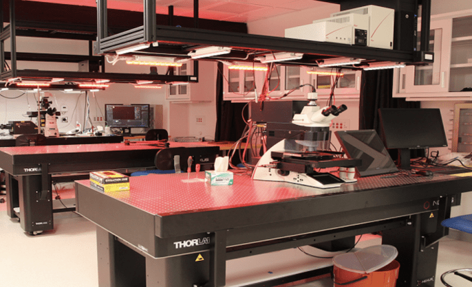
West Campus Imaging Core
We offer access to and training in the use of a variety of confocal and epifluorescence microscopes as well as light-sheet, laser microdissection, and atomic force microscopy.