Cores Directory
Search for the resources you need to answer your research questions.
Active Filters:
Listing of research core items
-
Core/facility
Yale Center for Molecular Discovery
We provide resources for assay development and high-throughput screening with small molecules and arrayed siRNA and sgRNA/CRISPR libraries in biochemical, cell-based and image-based assays (primarily 384-well format).
-
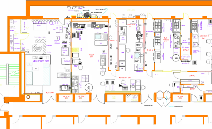
Core/facility
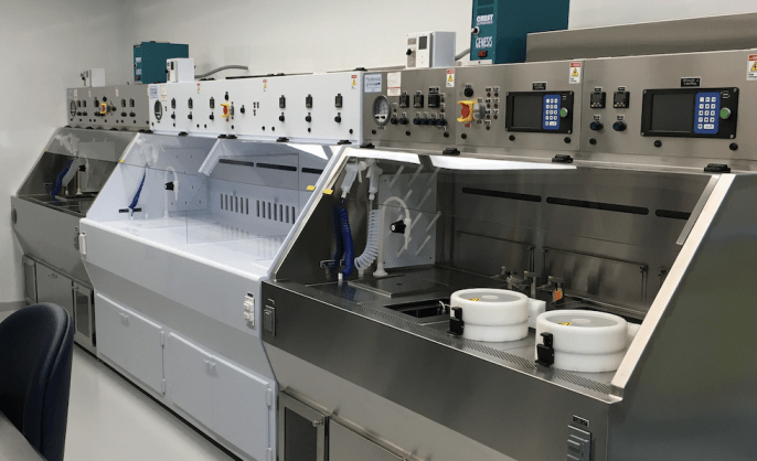
Yale University Cleanroom - West Campus
The cleanroom at West Campus includes 1200 square feet of wet and dry clean laboratories rated at Class 1000, with features standard to most cleanrooms.
-
Core/facility
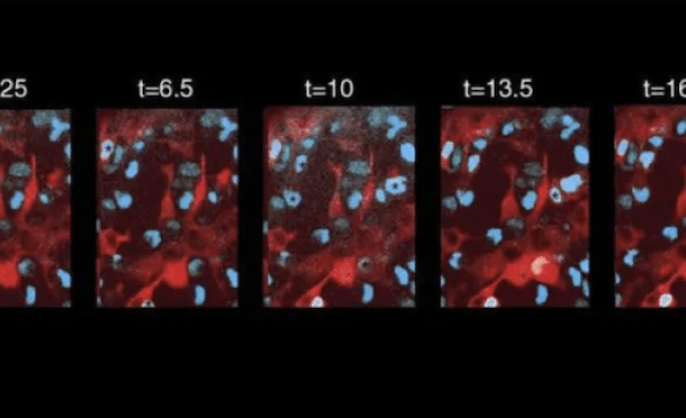
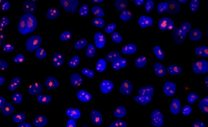
Confocal Microscopy at CCMI
We offer confocal microscopy, two-photon microscopy, light-sheet microscopy, swept-field microscopy, super-resolution imaging, and image analysis services.
-
Core/facility
Yale CryoEM Resource
The Yale CryoEM Resource comprises electron microscopes on all 3 campuses: a Titan Krios on West Campus, a Glacios and a Tecnai T12 at Yale School of Medicine, and a Glacios and a Talos L120C on Science Hill.
-
Core/facility
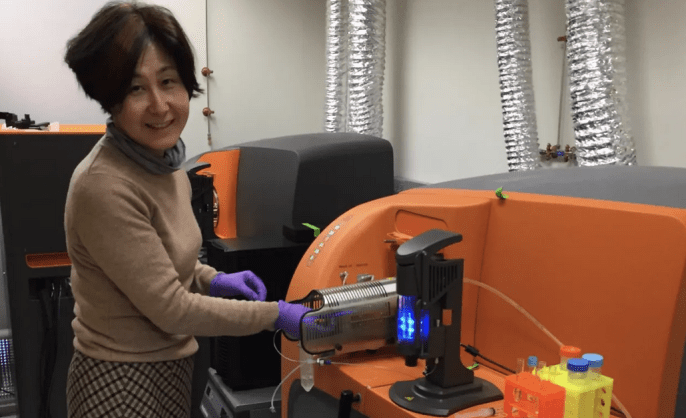
CyTOF Facility
The CyTOF 2 facility houses a time-of-flight atomic mass spectrometer for the high-speed acquisition of highly multi-parametric single-cell data.
-
Instrument/equipment
Dry and plasma etching
Oxford 100 RIE-ICP and Glow Research AutoGlow
Located within Yale University Cleanroom - West Campus -
Core/facility
Earth Materials Characterization Center (EMC2)
We offer materials physics equipment including microscopy, spectroscopy, deformation tools, and sample-prep tools for sample characterization work.
-
Core/facility
Electron Microscopy Facility at CCMI
The Electron Microscopy Facility at the Center for Cellular and Molecular Imaging (CCMI) provides investigators with a wide range of electron microscopy services.
-
Instrument/equipment
EM CCMI instruments
Microscopes, sample preparation, carbon evaporators, and advanced cryo FIB lamella milling for cryo electron tomography.
Located within Electron Microscopy Facility at CCMI -
Services
EM CCMI services
Project consultation and experimental design, sample preparation, and imaging with transmission and scanning electron microscopes.
Located within Electron Microscopy Facility at CCMI -
Core/facility
Cores pages FAQ for directors
Learn to create and edit your core's website on Research at Yale. This page is both a FAQ and a demonstration.
-
Core/facility
FIB-SEM Collaboration Core (F-SCC)
We utilize unparalleled enhanced FIB-SEM pipeline to generate large-volume three-dimensional electron microscopy data with nanometer isotropic resolution. Such data reveal critical insights about structure-function relationships in biology.