Structural Science Facility
KCL 7, 11 and SCL 26
Diffraction, Scattering and Imaging
Structural Science is an interdisciplinary field that brings together powerful techniques, including single-crystal X-ray diffraction (SCXRD) and powder X-ray diffraction (pXRD), to uncover the structural foundations of materials at unprecedented depth. This facility further expands access to advanced methodologies such as Microcrystal Electron Diffraction (MicroED), small- and wide-angle X-ray scattering (SAXS/WAXS), and micro-computed tomography (microCT). Our instrumentation is designed to directly probe real and reciprocal space across length scales, from the atomic to the mesoscale.
Small molecule X-ray policy
- Staff mounted samples will be mounted at the convenience of their schedule.
- Samples can be submitted on the table outside KCL 3.
- To independently use the single crystal diffractometers, you must have met one of the following subpoints:
- Completed CHEM 511 and 512.
- Completed an equivalent course/training at a different institution with a recommendation from the instructor/instrument manager.
- Independent access enables X-ray facility use for your own research.
- Independent use does not enable facility use for you to collect data on behalf of other researchers at Yale.
- Exceptions are granted on a case-by-case basis.
Samples can be submitted on the table outside KCL 3.
Available to Yale researchers & external researchers
Core websiteRates
2024/2025 External for-profit user rates
| Name | Inst. Number | Price ($) / Unit | Unit | Rate ($) / hour |
|---|---|---|---|---|
| X-ray | ||||
| X-ray single crystal screening | 39 | 75.00 | sample | n/a |
| X-ray data collection | 15 | 618.00 | dataset | n/a |
| X-ray data processing and structure report | 18 | 639.52 | structure | n/a |
| Powder diffraction | 14 | 75 | hour | 75 |
| MicroCT | 6 | 222.09 | hour | 222.09 |
| SAXSWAXS | 17 | 309.00 | hour | 309.00 |
| Electron Diffraction (MicroED/3DED) | ||||
| Sample preparation | 36 | 2060.00 | sample | n/a |
| Data collection | 37 | 2060.00 | dataset | n/a |
| Data processing and structure report | 38 | 2060.00 | structure | n/a |
| Hourly data collection | 40 | 250.00 | hour | 250.00 |
Structural Science Instruments
Learn more about our instruments below:
Pilatus3R HPC on a 007 VHF+Arc)Sec X-ray Source
Rigaku CCD on 007HF+ X-ray Source
Rigaku CCD on 007HF+Arc)Sec X-ray Source
Rigaku MiniFlex600 Powder X-ray Diffractometer
Rigaku XtaLAB Mini II with a hybrid pixel detector
Rigaku XtaLAB Synergy, Dualflex, HyPix-Arc 100
TTP Lab-tech Nanoliter Pipetting System
Xenocs Xeuss 3.0 Small/Wide X-ray Scattering System
XT H 225 Computed Tomography Scanner
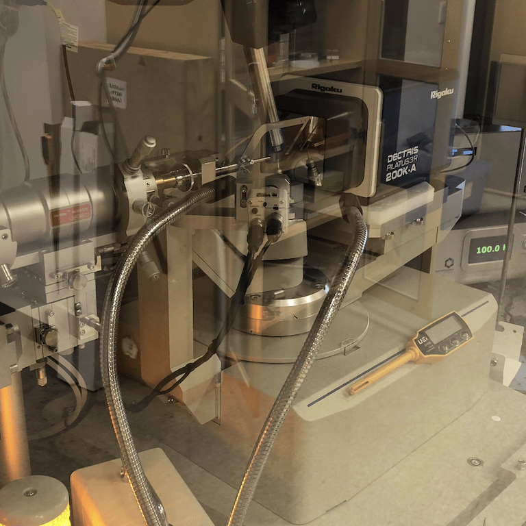
Short name: 007c
Pilatus3R HPC on a 007 VHF+Arc)Sec X-ray Source
Instrument location: KCL 7
Date of acquisition: October 2016
The Pilatus3R 200KA detector on our Rigaku 007 HF+ rotating anode with VHF Arc)Sec optics is suitable for solving small molecules samples. This instrument is run by the CBIC Staff. λ = 0.7107 Å
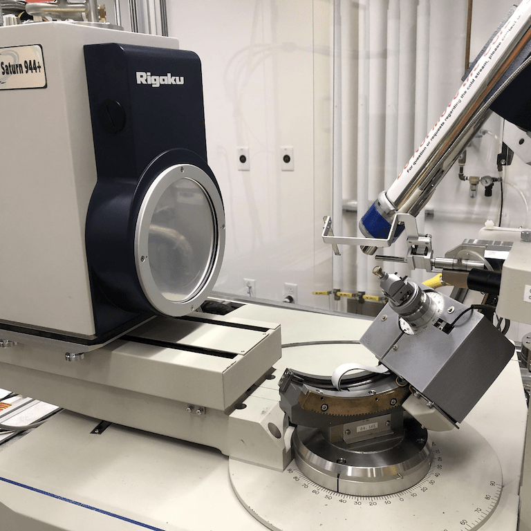
Short name: 007a
Rigaku CCD on 007HF+ X-ray Source
Instrument location: KCL 7
Date of acquisition: October 2009
The Saturn 944+ CCD detector on our Rigaku 007 HF+ rotating anode X-ray source with HF optics is suitable for solving small-molecule and protein structures by single-crystal X-ray diffraction. This instrument can also be used to screen crystals before sending them to an x-ray beam line at a synchrotron. λ = 1.5418 Å
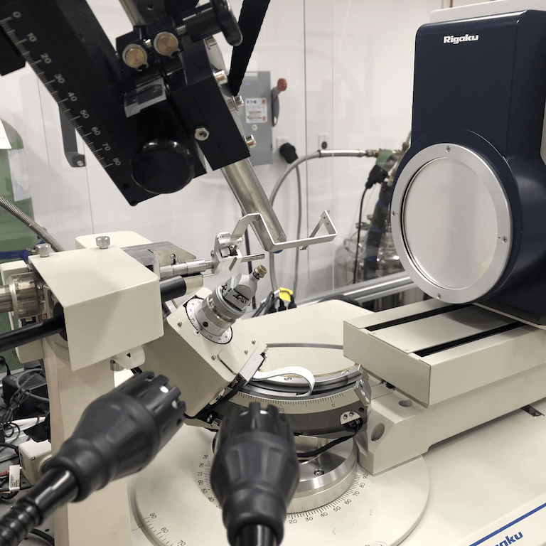
Short name: 007b
Rigaku CCD on 007HF+Arc)Sec X-ray Source
Instrument location: KCL 7
Date of acquisition: June 2016
The Rigaku Saturn 944HG CCD detector on our Rigaku 007 HF+ rotating anode X-ray source with HF Arc)Sec optics is suitable for solving small molecules samples. This instrument is run by the CBIC Staff. λ = 1.5418 Å
Short name: PXRD
Rigaku MiniFlex600 Powder X-ray Diffractometer
Instrument location: KCL 7
Date of acquisition: December 2004
This powder diffractometer can characterize crystallinity, crystal phases, and, in many cases, the identity of solid samples. Our instrument has a sealed-tube copper X-ray source, a scintillation counter with high dynamic range, and Bragg-Brentano geometry with slits providing high resolution for flat powder samples. λ = 1.5418 Å
Short name: mini2
Rigaku XtaLAB Mini II with a hybrid pixel detector
Instrument location: KCL 7
Date of acquisition: May 2022
The XtaLabMini II is a fully functional benchtop single-crystal X-ray diffractometer with a CCD detector. λ = 0.7107 Å
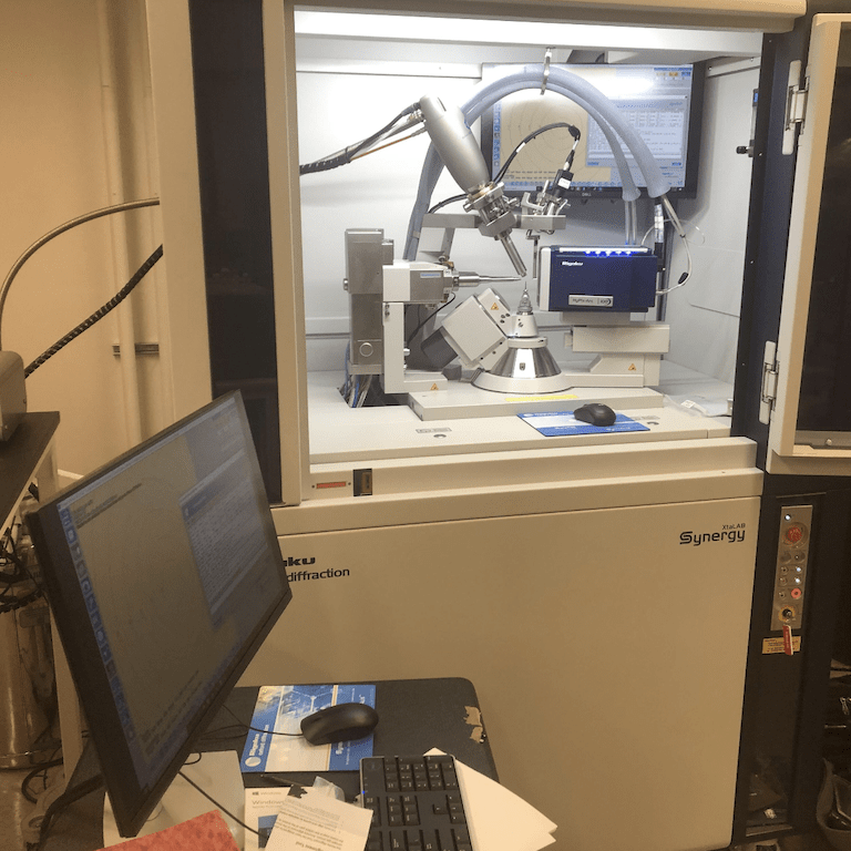
Short name: syn
Rigaku XtaLAB Synergy, Dualflex, HyPix-Arc 100
Instrument location: KCL 7
Date of acquisition: January 2023
Copper and silver X-ray sources are available. The curved image plate detector allows high dynamic range and collection out to a 2Θ angle of 144 degrees in a single image to yield high-resolution single-crystal data.
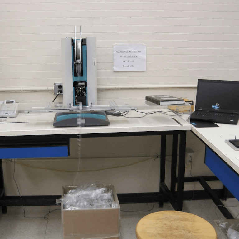
Short name: Mosquito
TTP Lab-tech Nanoliter Pipetting System
Instrument location: KCL 7
Date of acquisition: October 2009
TTP Labtech’s mosquito allows for the creation of protein crystallization screens with several multi-component drops per well, even in 96-well hanging drop set-ups.
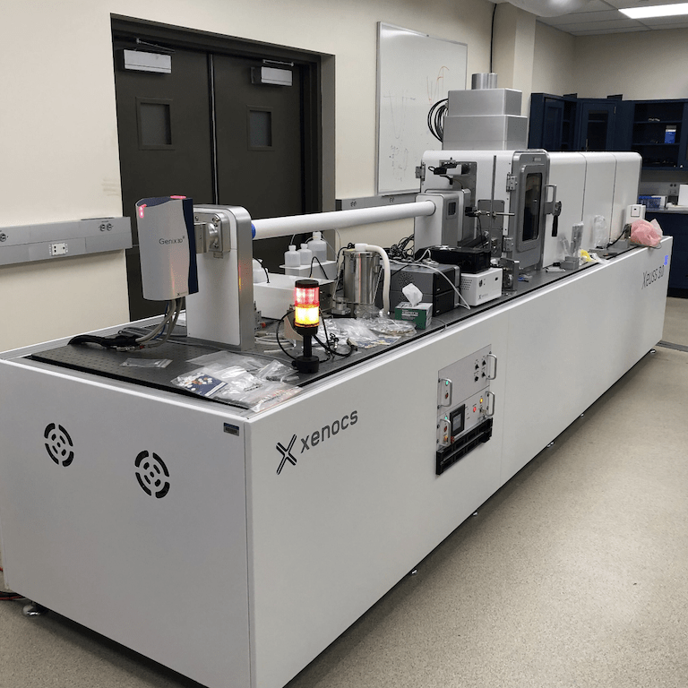
Short name: SAXSWAXS
Xenocs Xeuss 3.0 Small/Wide X-ray Scattering System
Instrument location: SCL 26
Date of acquisition: December 2022
The Xeuss 3.0 offers a maximum flexibility of measurement configurations to get the best possible data quality on any type of sample. In particular, the following key features enable the user to optimize experiments or results:
- High flux settings adapted for fast kinetics embedded in a low background camera
- Largest surface of detection moveable all the way from WAXS to long-distance SAXS to optimize resolution and signal-to-noise
- Long distance SAXS settings for measuring large characteristic dimensions (up to 900 nm depending on the Xeuss 3.0 model)
- Optional USAXS module to characterize large structures ( >2.5 µm)
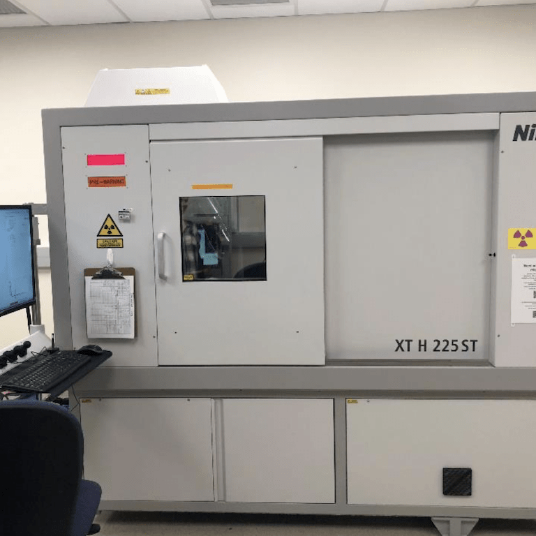
Short name: microCT
XT H 225 Computed Tomography Scanner
Instrument location: SCL 26
Date of acquisition: May 2018
Micro-CT is a 3D imaging technique utilizing X-rays to see inside an object, slice by slice. Micro-CT, also called microtomography or micro computed tomography, is similar to hospital CT or “CAT” scan imaging but on a small scale with greatly increased resolution. Samples can be imaged with pixel sizes as small as ~1 micron and objects can be scanned up to ~50 cm in diameter.
The versatile XT H 225 scanner can be used to cover a wide range of applications, including the inspection of plastic parts, electronics and complex mechanisms as well as researching materials and natural specimens.
The scanner can be fitted with a rotating anode, reflection, or transmission target. The rotating anode produces the most X-rays, which is good for highly absorbing material, but has the lowest resolution. The transmission target has the best resolution, but produces the least amount of X-rays. The reflection target is typically installed and has a balance between X-ray intensity and resolution. If you need the rotating or transmission target, please inquire in advance about scheduling.
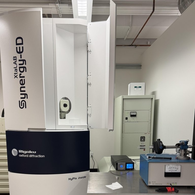
XtaLab Synergy-ED
The Synergy-ED is a state-of-the-art electron diffractometer designed for Microcrystal Electron Diffraction (MicroED) / 3D Electron Diffraction (3DED) experiments. It enables high-resolution structural analysis of nanocrystalline materials that are too small for conventional X-ray diffraction techniques.
Key Features
Wavelength:
Operates at an electron wavelength of ~0.0251 Å with a 200 kV accelerating voltage, providing high-resolution diffraction data.
Temperature Range:
Capable of measuring samples from room temperature down to 100 K, allowing for studies of temperature-dependent structural changes.
Viable Grain Size:
Suitable for nanocrystals and microcrystals ranging from several hundred nanometers to less than 10 microns.
Sample Types
The Synergy-ED is optimized for a broad range of crystalline materials, including:
-Small organic molecules (e.g., pharmaceuticals, natural products)
-Inorganic nanomaterials
-Metal-organic frameworks (MOFs) and zeolites
-Peptides and small proteins
-Polymers and hybrid materials
Applications
Pharmaceutical Research:
Polymorph Studies: Determines different crystal forms of a compound, crucial for drug formulation and stability.
Phase Transitions: Investigates temperature- or pressure-induced structural changes.
Materials Science & Nanotechnology:
Grain Boundary Analysis: Characterizes defects, interfaces, and grain orientations in polycrystalline materials.
Nanocrystalline and Amorphous Phase Identification: Helps differentiate between ordered and disordered structures.
Heterogeneous Sample Analysis:
Sample Mixtures: Can resolve and identify multiple crystalline phases within complex mixtures.
Solid-State Chemistry: Provides insight into phase purity and composition.