Listing of research core items
-
Instrument/equipment
Functional near-infrared spectroscopy (fNIRS)
Functional near-infrared spectroscopy (fNIRS) is a portable technique that uses near-infrared light to estimate cortical hemodynamic activity related to neural activity.
Located within BrainWorks -
Services
Gas chromatography
Measure greenhouse gas concentrations.
Located within Yale Analytical and Stable Isotope Center (YASIC) -
Core/facility
Yale Genome Editing Center
We create genetically engineered mice via CRISPR/Cas, gene targeting via homologous recombination in ES cells, and traditional transgenic technology. We also cryopreserve mouse sperm and embryos.
Timothy Nottoli, PhD
Co-Director Research Scientist in Comparative Medicine -
Services

Glass shop products
Examples of our offerings include adapters, bubblers, bump traps, chromatography columns, condensers, dry-storage jars, filtration funnels, flasks, Kjeldahl traps, Schlenk ware, and vacuum traps.
Located within Scientific Glassblowing Laboratory (Glass Shop)Daryl Smith
-
Instrument/equipment
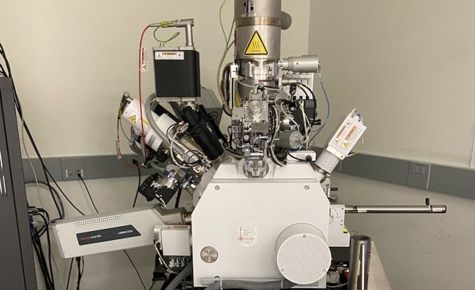
Helios G4 UX DualBeam System
Helios G4 UX DualBeam (electron & ion-beam) microscope
Located within Aberration-Corrected Electron Microscopy (ACEM) Core -
Services
Histology Service
Preparation of animal tissues for microscopic examination.
Located within Comparative Pathology Research Core -
Instrument/equipment
Hitachi IM 4000 ion milling
The IM4000II supports both cross-section milling and flat-milling to prepare specimens, depending on the purpose.
Located within Earth Materials Characterization Center (EMC2) -
Services
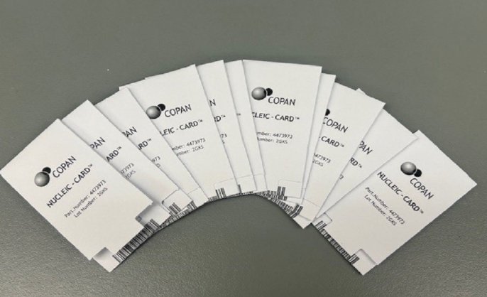
Human cell line authentication
Using a cell line authentication service can help to establish the identity of a cell line or confirm that it remains uncontaminated. With the use of the CLA Identifiler Direct Kit, we will perform STR genotyping by fragment analysis. This STR profile will be the key to authenticating the cell line.
Located within Keck DNA Sequencing Facilitydnaseq@yale.edu
-
Core/facility
Human Embryonic/Induced Pluripotent Stem Cell Core
We provide essential services for research on human embryonic stem cells (hESCs) and human induced pluripotent stem cells (iPSCs).
-
Services
Imaging services
Micro-CT, pulse Doppler, and imaging analysis.
Located within Yale CVRC Micro-CT Imaging CoreZhen Zhuang, MD, MS
-
Instrument/equipment
Imaris image analysis workstations
Imaris imaging analysis workstations
Located within Confocal Microscopy at CCMI -
Services
In vitro assays and cancer models
3D and explant cultures, cytotoxicity assays, and immunoassays.
Located within Precision Cancer ModelingVish Muthusamy, PhD