Magnetic resonance imaging (MRI)
Building: 100 College St
About the instrument
MRI reveals the structure and function of the brain at high spatial resolution. The MRI suite is equipped with a full range of stimulus delivery and response devices and a mock scanner for pretraining and acclimation to the MRI environment.
Available to Yale researchers only
Specifications
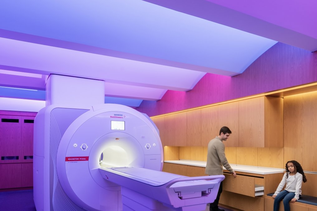
Siemens MAGNETOM Prisma 3T scanner
- 3-Tesla magnetic field
- 20-channel, 32-channel, and 64-channel head and head/neck coils
- Maximum amplitude of 80 mT/m @ a slew rate of 200 T/m/s
- Sequences for rapid, high-resolution functional, structural, diffusion weighted, susceptibility weighted, and quantitative imaging
Stimulus and response peripherals
- OptoAcoustics OptoActive III adult and newborn headset with active noise cancellation for high-fidelity audio delivery at high frequencies
- Sensimetrics S15 in-ear audio
- OptoAcoustics fiber optic microphone with active noise cancellation
- PST Hyperion projector system, 1080p@60 Hz
- SR Research EyeLink 1000+ eye tracker
- BIOPAC MP160 with EDA module
- A range of fiber optic response boxes
Mock scanner
- PST Vera MRI simulator
- Realistic 60 cm bore and 32-channel head coil
- Real-time head movement measurement
- Simulates the acoustic environment of the MRI
- Screen and trainer response boxes
Rates
$400/hr
Resources and documentation
Training & services
Users are responsible for safe operation of the equipment and data collection. Two people must be present for data collection. All users must read the user manual and complete an in-person orientation. Unsupervised operation is permitted after passing five consecutive supervised sessions. Please request training here.