Analysis: Amnis Imagestream-X MarkII Imaging Flow Cytometer
Location & contact — Table — Optics — Links
Amnis Imagestream-X MarkII imaging flow cytometer provides users with the ability to gain detailed images of a large number of cells in a relatively short period of time and with the opportunity to perform a range of novel applications including co-localization, internalization, stem cell differentiation, and cell-cell interactions.
ImageStreamX MarkII produces up to 12 high resolution images of each cell directly in flow, at rates exceeding 1,000 cells per second, and with the fluorescence sensitivity of conventional flow cytometers. These capabilities allow you to quantitate cellular morphology and the intensity and location of fluorescent probes on, in, or between cells, even in rare sub-populations and highly heterogeneous samples.
With the ImageStreamX MarkII you can:
- Image cells directly in suspension with the resolution of 20X, 40X or 60X objective and the fluorescence sensitivity of flow cytometers
- Analyze highly heterogeneous samples and rare cell sub-populations at speeds exceeding 1,000 cells per second
- Perform phenotypic and functional studies at the same time using up to five lasers and 12 images per cell
- Quantitate virtually anything you can see using the IDEAS® software package’s numerous pre-defined fluorescence and morphologic parameters
Location & contact
Location: The Anlyan Center (TAC) S613
Contact: Ashraf Khalil, 203-785-7958
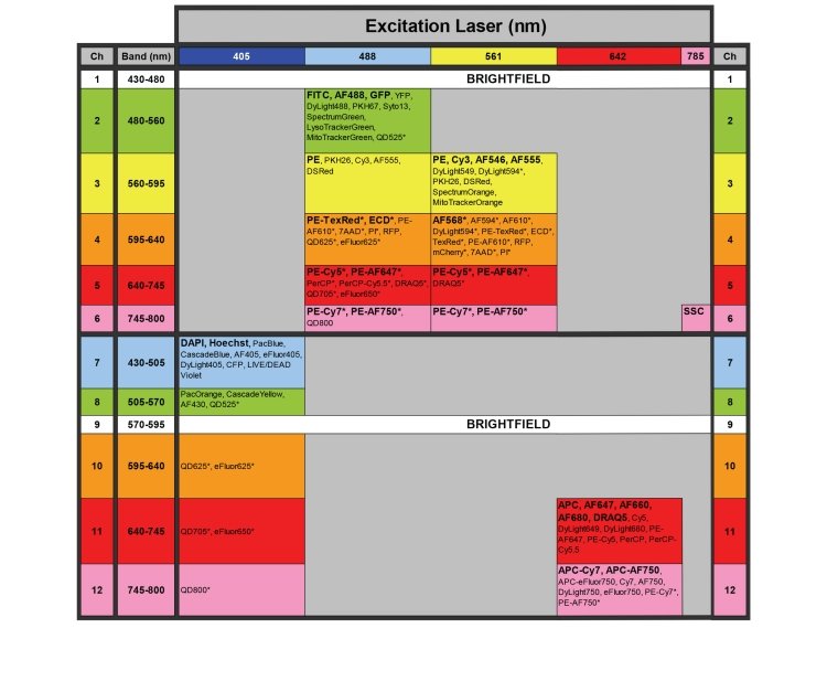
Table

Or view the above chart in PDF form.
Optics
ImageStreamX has 4 lasers (405 nm violet laser, 488 nm blue laser, 561 nm green laser, 642 nm red laser and a 785 nm laser used by SSC).
The output of each laser can be independently adjusted for greater experimental flexibility and optimal results.