Cores Directory
Search for the resources you need to answer your research questions.
Active filters:
Listing of research core items
-
Instrument/equipment
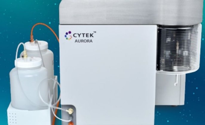
Cytek Aurora Spectral cell analyzer
Analyze from single tubes, 40-tube racks, or 96-well plates with 5 lasers (UV, violet, blue, yellow/green, red) and 64 detectors (16 UV, 16 violet, 14 blue, 10 yellow/green, 8 red).
Located within West Campus Flow Cytometry Facility -
Instrument/equipment
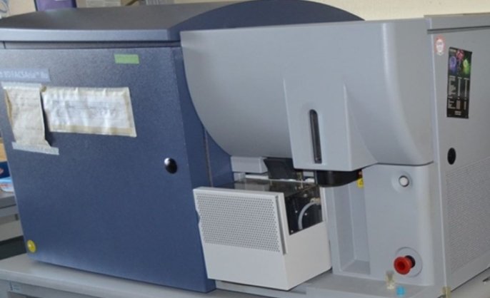
BD FACS Aria cell sorter
Sort into 5 mL round tubes, 15 mL conical tubes, or plates (24-, 48-, or 96-well) with 4 lasers (violet, blue, yellow/green, red) and 14 detectors (6 violet, 2 blue, 4 yellow/green, 2 red).
Located within West Campus Flow Cytometry Facility -
Instrument/equipment

FEG Electron Microprobe
This FEG electron microprobe can acquire high-resolution SEI and BSE images in support of qualitative, semi-quantitative, and quantitative-WDS microscale chemical analysis.
Located within Earth Materials Characterization Center (EMC2)Jim Eckert, PhD
-
Instrument/equipment
FEMTO3D Atlas super high-speed two-photon microscope
Flexible, high-speed imaging system using acousto-optical deflectors; capable of capturing activity from over 2,000 neurons or dendritic structures in parallel in vivo.
Located within Wu Tsai Institute Microscopy Shared FacilityWTI Microscopy
-
Instrument/equipment
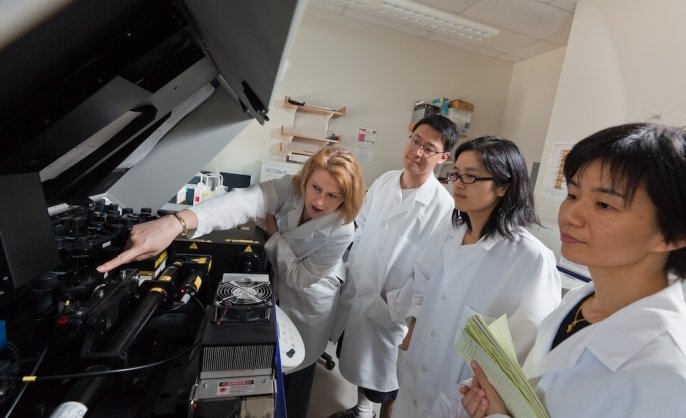
Flow cytometry analysis
We are equipped with user-operated analyzers at three different locations. We are open to all researchers throughout Yale. New users should create an account in PPMS and have their business office authorize financial information.
Located within Yale Flow Cytometry Facility -
Instrument/equipment
Functional near-infrared spectroscopy (fNIRS)
Functional near-infrared spectroscopy (fNIRS) is a portable technique that uses near-infrared light to estimate cortical hemodynamic activity related to neural activity.
Located within BrainWorks -
Instrument/equipment
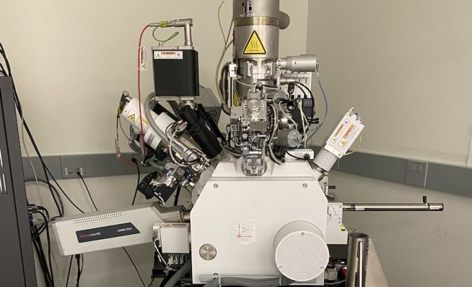
Helios G4 UX DualBeam System
Helios G4 UX DualBeam (electron & ion-beam) microscope
Located within Aberration-Corrected Electron Microscopy (ACEM) Core -
Instrument/equipment
Hitachi IM 4000 ion milling
The IM4000II supports both cross-section milling and flat-milling to prepare specimens, depending on the purpose.
Located within Earth Materials Characterization Center (EMC2) -
Instrument/equipment
Imaris image analysis workstations
Imaris imaging analysis workstations
Located within Confocal Microscopy at CCMI -
Instrument/equipment
In vivo imaging
The PCM provides user access to in vivo imaging instruments housed within imaging clusters at two campus locations. These instruments are available for independent user operation following completion of required instrument and regulatory training.
Located within Precision Cancer ModelingVish Muthusamy, PhD
-
Instrument/equipment
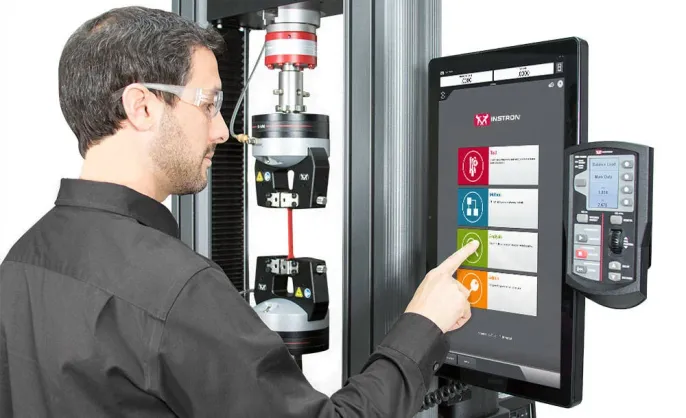
Instron tensile/compression testing
This instrument performs tensile, compression, flexure/bend, peel, tear, shear, and cyclic tests.
Located within Mechanical and Thermal Analysis Instrumentation Core (MTAIC) -
Instrument/equipment
Instruments for sample preparation
The core houses multiple cryo-sample preparation devices.
Located within Yale CryoEM Resource