Developmental Histology/Tissue Microarray & Archiving
About the services
Developmental Histology/Tissue Microarray is a shared resource of the Yale Cancer Center & Yale Pathology Tissue Services.
Mission
To provide:
- Timely high-quality histology services to the Yale community and beyond.
- Access to tissue services from the Yale Pathology tissue archives.
- To maximize the value of the resources in the Yale Pathology Tissue Archive.
- To provide tissue microarrays to assess multiple tissue samples.
Services
Tissue Microarray & Archiving
We provide the university community with services related to tissue microarray technologies. Services are also available for the biotech and pharmaceutical industries.
These services include:
- Array design and construction
- Slide distribution: master and test array slides
- Technical support
- Cell line and tissue controls provided
Histology
Our histology services include:
- Special stains
- Immunohistochemistry
- IHC workup
- Frozen sectioning
Available to Yale researchers only
What are tissue microarrays?
Tissue microarrays are produced by a method of re-locating tissue from conventional histologic paraffin blocks such that tissue from multiple patients or blocks can be seen on the same slide. This is done by using a needle to biopsy a standard histologic sections and placing the core into an array on a recipient paraffin block. This technique, originally described by in 1987 by Wan, Fortuna and Furmanski in Journal of Immunological Methods. They published a modification of Battifora's "sausage" block technique whereby tissue cores were placed in specific spatially fixed positions in a block. The technique was popularized by Kononen and colleagues in the laboratory of Ollie Kallioneimi after a publication in Nature Medicine in 1998. This technology should not be confused with DNA microarrays where each tiny spot represents a unique cloned cDNA or oligonucleotide. In tissue microarrays, the spots are larger and contain small histologic sections from unique tissues or tumors.
Example
The arrays are assembled by taking core needle “biopsies” from specific locations in pre-existing paraffin-embedded tissue blocks and re-embedding them in an arrayed “master” block, using techniques and an apparatus developed by Konenen et al. In this way, tissue from 600 specimens can be represented in a single paraffin block.
Construction

The arraying device is designed to produce sample circular spots that are 0.6 mm in diameter at a spacing of 0.7-0.8 mm. The surface area of each sample is 0.282 mm2, or in pathologists’ terms, about the size of 2-3 high power fields. The number of spots on a single slide is variable depending on the array design, the current comfortable maximum with the 0.6 mm needle is about 600 spots per standard glass microscope slide.
Quantitative analysis

Once a TMA is constructed it can be used for quantitative analysis. AQUA technology measures the actual level of protein in the cells by fluorescence. This is a much more sensitive and specific way to measure proteins for more specialized chemotherapeutic treatment.
Histology services
Special stains
Our special stains include, but are not limited to:
- Alcian blue
- Alizarin red
- Congo red
- D.Pas
- EVG
- Mucicarmine
- Movat
- Oil red O.
- Reticulin
- Sirius red
Download a list of all the stains we currently provide.
Immunohistochemistry
Services include working up antibodies for research utilizing animal models.
We also provide and work up antibodies for human tissue use.
We provide antibodies from our standard list (provided upon request) plus the cost of IHC staining.
Antibodies, provided by the client, can be worked up through our services plus the cost of IHC staining.
IHC workup
Frozen sectioning
Ordering
eHistoEmbedding instructions
The diagrams illustrate a great way for you to describe how you would like your tissue embedded.
NOTE: We do NOT perform necropsy!


Specimen handling instructions
After placing an order online, print the order PDF to bring to the YPTS labs at CB-562 along with your specimen(s). Follow the below instructions for specimen submission.
- Fix specimens for 12-24 hours. Some may require greater fixation time based on tissue size.
- Transfer specimens into 70% alcohol. This is recommended for long-term storage and required for transport. For the detection of fat please submit a frozen specimen in OTS.
- Please clearly label containers that contain neutral buffered formalin or PFA. Specimens should have an outer container when being transported. This is a critical requirement for specimen handing and transport.
- Label cassettes in pencil only. Ink washed off during processing. Developmental Histology is not responsible for cassettes labeled in ink.
- Minimal writing on cassettes (must be legible). The cassette should be labeled with the experiment number, the animal number, and/or organ or site and/or "A", "B", "C", or "1", "2", "3". The slide label software can only hold a few lines of text. We will only label the slide with the information that will fit per our software that appears on the cassette.
- Novel materials: Developmental Histology is not responsible for the outcome of any "novel" antibody, tissue, or materials submitted for workup. The customer will be charged according to our fee schedule for tech time and efforts.
- Include a Word or Excel document of specimens' designations when dropping off your specimens. This is valuable if the containers or cassettes are partially illegible.
- Your orders will be available online for editing until they are marked as verified in our system. Order status can be checked throughout its time in the lab in the online ordering system at any time. Previous orders in the new system can also be viewed and are part of your account information.
- Deadlines: If your order is needed by a certain deadline, please let us know. We do orders as they are received/verified in the lab.
Make sure that your tissue:
- Does not touch the edge of the cassette
- Is not too thick
NEVER squeeze a tissue sample into a cassette.

Contacts
David L. Rimm, MD, PhD, Medical Director
Location: CB-568
Tel: 203-737-4198
Tel: 203-785-5879
Fax: 203-737-1198
Mailing:
Yale School of Medicine
Department of Pathology
PO Box 208023
New Haven, CT 06520-8023
To ship specimens:
Yale School of Medicine
Department of Pathology
310 Cedar St., CB-568
New Haven, CT 06520
-
Services
Cellular and mitochondrial respiration and bioenergetics
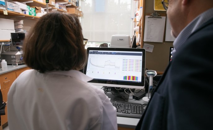
Cellular and mitochondrial respiration (OCR) and acidification (ECAR) are assessed with the Agilent Seahorse XF Pro 96. In a 96-well plate format, real-time measurements of cellular respiration, oxidative phosphorylation, mitochondrial leak, and mitochondrial transport are assessed as functional kinetic measurements of cellular bioenergetics. Changes in acidification are also observed simultaneously. An integrated Agilent BioTek Cytation5 imager correlates cell number via Hoechst nuclear stain to respiration for more accurate normalization of respiration.
Located within Chemical Metabolism CoreRebecca Cardone, MS
-
Instrument/equipment
BD LSR Fortessa X20
The Fortessa analyzer has 4 lasers, including V 405 nm, B 488 nm, YG 561 nm, and R 640 nm. It can detect 16 colors.
Located within Science Hill Flow Cytometry FacilityFreya Wencker, PhD
-
Instrument/equipment

BD Symphony A1 analyzer
Analyze from single tubes or 96-well plates with 45 lasers (violet, blue, yellow/green, red) and 64 detectors (6 violet, 2 blue, 4 yellow/green, 3 red).
Located within West Campus Flow Cytometry Facility