Tissue Procurement and Distribution (TPD)
About the service
Tissue Procurement and Distribution (TPD) is a shared resource of Yale Cancer Center & Yale Pathology Tissue Services.
Mission
The mission of this division is to maximize the access of human tissues to translational investigators by providing high quality annotated fresh and frozen human tissue to Yale and other users.
Underlying premises
Available to Yale researchers only
Standard operating protocol
It is essential to perform optimal management of pathology resources including fresh, frozen and formalin-fixed specimen regarding the requests of human specimens for research purpose. All human tissues must be handled in strict compliance with HIPAA regulations, university HIC policies, and diagnostic requirements. All users must recognize and be prepared to assume the responsibilities associated with all aspects of tissue procurement and management.
For each request, a standard operating procedure (SOP) must be drawn up by the requesting investigator and TPD director. The detailed condition and specific guideline of specimen grossing required should be defined by requesting investigator and attending pathologist in an effect to standardize operating and to keep research tissue sampling under management without jeopardizing diagnosis tissue submission.
Personnel from YTPDF will help interested investigators design and optimize SOPs. Terms for specific requests of specimens will be discussed depending on demand, specimen site, and the amount of Pathology participation and manpower required. Negotiations will be conducted by the director of Yale Pathology Tissue Services and overseen and approved by the Tissue Resource Oversight Committee (TROC).
For investigators whose research projects are not claimed to involve human subjects, based on institutional HIC guidelines, limited information associated with samples will be provided including age range, sex, diagnosis and pathological status (Tumor or Normal). A YTPS TPD party will assign a new identifier for each SOP-specific sample, in such a way that it would not be possible for investigators to identify the corresponding patient-subject directly. For linked tissues, investigators are required to have HIC approval and signed patient consent.
Prior to implementation, drafted SOP must be reviewed and approved by the directors of Pathology service and other appropriate individuals including attending pathologist who supervising specified tissue collection. Implementation date should be recorded for all procedures.
All data related to tissue collection must be entered into the YTPS TPD database, which will allow YPTS to manage, keep track of YPTS fresh and frozen tissue biorepositories, and to oversight and coordinate inventories in the distributed banks.
YTPS TPD SOP format
A. Purpose or clinical significance
B. Forms and records
C. Conditions for patient preparation
D. Step-by-step details of the procedure (detailed and specific):
- Collection personnel
- Collection site
- Collection logistics
- Exclusion and inclusion criteria for potentially legitimate specimens
- Grossing guideline for both tumorous and normal specimen sampling
- Anticipated ischemic time
- Minimal number of tissue unit required
- Minimal size (mm) required for each tissue unit
- Tissue processing
- Subjectivity to embargo period
- Tissue distribution
E. Equipment, reagents, supplies and personnel
F. Hazardous materials Material Safety Data Sheet (MSDS)
G. Distribution and location of SOP
Request tissue
Tissue request formEmbargo & patient safety
To prevent tissues from being used in a manner that jeopardizes patient care, an embargo policy is in place. This means tissue must be held in storage prior to research use for a time period dependent on tissue type, as outlined below.
| YPTS embargo timetable: Tissue type | Time |
|---|---|
| Frozen tissue | One month after sign-out |
| Fresh tissue (for cell culture) | None |
| Formalin-fixed paraffin-embedded tissue | One year from accession date |
Reference materials
Supplies
- Liquid nitrogen in appropriate LN2 transport container
- Isopentane stored at room temperature in a flammable cabinet
- RNAlater
- Tissue Tec OCT cryo-compound at room temperature
- Tissue cryomolds
- 10% neutral-buffered formalin
- Saline
- RPMI Medium 1640, used for the culture of human normal and neoplastic leukocytes
- PBS (phosphate-buffered saline)
Processing methods
- Flash freezing
- Freezing by direct immersion in liquid nitrogen is common, although it is more damaging to the tissue. Rapid freezing in isopentane results in less tissue damage.
- Isopentane is an extremely volatile and flammable liquid at room temperature and pressure.
- Vapor or mist is irritating to the eyes, skin, mucous membrane, and upper respiratory tract. Precaution should be taken to avoid contact to the skin and other body surface.
- Pour approximately 100 to 200 ml isopentane into a plastic beaker. Place the beaker into liquid nitrogen or dry ice. Isopentane can be cooled to its freezing point (–160°C).
- The pre-labeled cassette with sample will be placed in isopentane cooled by dry ice or liquid nitrogen. Do not remove the tissue from isopentane until freezing is complete (10 seconds or less depending on size) but ensure sample does not crack.
- Isopentane disposal method: allow gas to vent slowly to atmosphere in an unconfined area or exhaust hood.
- Remain in RNAlater
- As specific and non-specific RNA degradation begins immediately after tissue harvest, prompt transfer of fresh specimens into an RNA stabilization reagent is helpful to minimize changes in gene expression, and to ensure accurate quantitative analysis of gene expression.
- RNAlater can be stored at room temperature. If any precipitation is seen, heat it to 37°C and agitate to redissolve. RNAlater can safely be discarded down the sink and flushed with water.
- Tissue stored in RNAlater is initially kept overnight at 2–8°C (minimum 12 hours).
- After incubation in RNAlater at 4°C overnight, the following options are available until processing begins:
- Samples can remain in RNAlater for up to 1 day at 37°C, 7 days at 18–25°C, or 4 weeks at 2–8°C, allowing processing, transportation, storage, and shipping of samples without liquid nitrogen or dry ice.
- Alternatively, the samples can also be placed at –20°C or –80°C for archival storage (see Reference 1).
- OCT embedding
- OCT protocol is used for freezing tissue samples when good preservation of histological details is required. OCT samples can also be used for protein, DNA and RNA extraction if there is enough tissue.
- Put drops of OCT into a plastic cryomolds. Place tissue on top in correct orientation for cutting. Then carefully pour OCT on top of tissues to make sure none of the tissues remain exposed. Bubbles should be avoided.
- Submerge cryomold with OCT embedded sample into isopentane cooled by dry ice or liquid nitrogen until OCT has hardened. OCT specimens will be subsequently placed either in –80°C freezer or in liquid nitrogen tank for storage.
- Keeping tissue fresh for subsequent cell culture
- Separate blades or scalpels or razor blades for bisecting samples should be used for each anatomic site to avoid cross-contamination.
- Tissue sample should be placed into 15 ml or 50 ml conical tube filled with at least 10 times of lab-made specific medium or the following reagents in a timely manner.
- RPMI Medium 1640, used for the culture of human normal and neoplastic leukocytes
- PBS (phosphate-buffered saline)
- Keep the tissue at 4°C until tissue distribution.
- 10% Formalin fixation
- 10% Neutral-buffered formalin (10% NBF) contains 4% formaldehyde and is the most commonly used fixative in pathology.
- It is toxic by inhalation and if swallowed, can cause irritation to eyes, the respiratory system and the skin. Precaution should be taken to avoid inhalation, and contact the skin and other body surface.
- Formalin should not be discarded down the drain. Refer to the chemical disposal plan for appropriate method of disposal.
- Tissue fixation in formalin
- Tissue should be no bigger than 20 x 10 x 5 mm.
- The volume of formalin should be a minimum of 15 times the volume of the tissue samples. Containers should be capped, leak-proofed.
- The advantage of formalin fixation is that tissue morphology is maximally preserved.
- The most common molecular change induced by formalin fixation is the formation of cross-links between proteins, or between proteins and nucleic acids. This may be responsible for the masking the epitope by altering the three-dimensional structure of the protein. Enzyme digestion or heat-induced epitope retrieval may rescue unavailable sites (see References 2, 3).
- Duration and timing of formalin fixation
- Tissue fixation time is dependent on tissue size. Small tissue piece (10 x 10 x 3 mm) fixed in 10% NBF for 6 to 24 hours will generally show good cytological preservation.
- Short formaldehyde fixation before tissue dehydration and embedding in paraffin.
- Short fixation results in cross-linking only at the periphery of the tissue block.
- Coagulation by alcohol during tissue dehydration does occur in the center of the tissue, or the center actually remains raw and not fixed. Intensity of immunostaining of center area will be varied when comparing the well-fixed periphery area.
- Prolonged formaldehyde fixation (longer than 48 hours)
- Prolonged formaldehyde fixation results in excessive cross-linkage, lead to weak or absent staining, though increasing antigen retrieval step may counteract the effect of prolonged formaldehyde fixation (see Reference 4).
- Delay of fixation
- Delayed fixation results in irreversible weak or absent staining resulted from protein dephosphorylation or protein degradation (see References 5 & 6).
Suggestion: If specimen cannot be fixed in a timely manner, refrigeration of the specimen may be helpful and is recommended.
- Delayed fixation results in irreversible weak or absent staining resulted from protein dephosphorylation or protein degradation (see References 5 & 6).
- Short formaldehyde fixation before tissue dehydration and embedding in paraffin.
- Tissue fixation time is dependent on tissue size. Small tissue piece (10 x 10 x 3 mm) fixed in 10% NBF for 6 to 24 hours will generally show good cytological preservation.
- Tissue fixation in formalin
References
- Florell SR, Coffin CM, Holden JA, et al. Preservation of RNA for functional genomic studies: a multidisciplinary tumor bank protocol. Mod Pathol 2001; 14(2):116-28.
- Helander KG. Kinetic studies of formaldehyde binding in tissue. Biotech Histochem 1994; 69(3):177-9.
- Fox CH, Johnson FB, Whiting J, Roller PP. Formaldehyde fixation. J Histochem Cytochem 1985; 33(8):845-53.
- Werner M, Chott A, Fabiano A, Battifora H. Effect of formalin tissue fixation and processing on immunohistochemistry. Am J Surg Pathol 2000; 24(7):1016-9.
- Bai Y, Tolles J, Cheng H, et al. Quantitative assessment shows loss of antigenic epitopes as a function of pre-analytic variables. Lab Invest 2011; 91(8):1253-61.
- Neumeister VM, Anagnostou V, Siddiqui S, et al. Quantitative assessment of effect of preanalytic cold ischemic time on protein expression in breast cancer tissues. J Natl Cancer Inst 2012; 104(23):1815-24.
Process flowchart
Flowchart highlighting steps in the TPD process.

PI fills out TPD request form → PI meets with TPD to specify request → SOP writing (by TPD director & PI) → SOP approval (by attending pathologist, directors of Surgical Pathology, Autopsy Service, TPD, and YPTS) → Tissue procurement (by TPD staff) → Tissue documenting (by TPD staff) → Tissue distribution (including corresponding de-identifing clinical information) (by TPD staff) → Tissue order charging (by TPD director)
Contacts
Anita Huttner, MD, Medical Director
Yalai Bai, PhD, MD, Research Scientist; Technical Director
Dongqing Liu, MD, MSc, Research Assistant 1 MS; Technician
Location: BML 112
Tel: 203.785.4205
Fax: 203.785.3583
Mailing:
Yale School of Medicine
Department of Pathology
PO Box 208023
New Haven, CT 06520-8023
To ship specimens:
Yale School of Medicine
Department of Pathology
310 Cedar St., BML 112
New Haven, CT 06520
-
Services
Cellular and mitochondrial respiration and bioenergetics
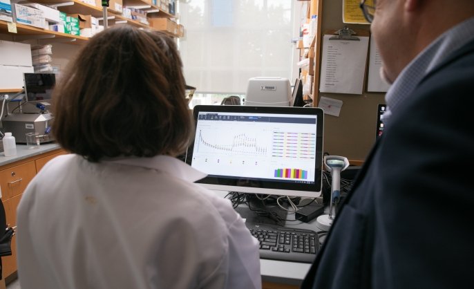
Cellular and mitochondrial respiration (OCR) and acidification (ECAR) are assessed with the Agilent Seahorse XF Pro 96. In a 96-well plate format, real-time measurements of cellular respiration, oxidative phosphorylation, mitochondrial leak, and mitochondrial transport are assessed as functional kinetic measurements of cellular bioenergetics. Changes in acidification are also observed simultaneously. An integrated Agilent BioTek Cytation5 imager correlates cell number via Hoechst nuclear stain to respiration for more accurate normalization of respiration.
Located within Chemical Metabolism CoreRebecca Cardone, MS
-
Instrument/equipment
BD LSR Fortessa X20
The Fortessa analyzer has 4 lasers, including V 405 nm, B 488 nm, YG 561 nm, and R 640 nm. It can detect 16 colors.
Located within Science Hill Flow Cytometry FacilityFreya Wencker, PhD
-
Instrument/equipment

BD Symphony A1 analyzer
Analyze from single tubes or 96-well plates with 45 lasers (violet, blue, yellow/green, red) and 64 detectors (6 violet, 2 blue, 4 yellow/green, 3 red).
Located within West Campus Flow Cytometry Facility