Confocal Microscopy at CCMI
We offer confocal microscopy, two-photon microscopy, light-sheet microscopy, swept-field microscopy, and super-resolution imaging services.
Sterling Hall of Medicine, room IE-25

About the core
Confocal microscopy enables the visualization and imaging of fixed as well as living cells and tissues that contain fluorescent probes (fluorescent antibodies, fluorescent proteins, dyes, and substrates). This technique allows sharply defined optical sections to be collected, from which three-dimensional rendering and movies can be created.
Our facility offers confocal microscopy, two-photon microscopy, light sheet microscopy, swept-field microscopy, and super-resolution imaging services to Yale investigators. It provides access to and training in the use of all microscopes, related equipment, and image workstations.
Equipment and services
- Leica SP5 confocal microscope
- Stellaris 8 DIVE multiphoton microscope
- Zeiss LSM 880 Airyscan confocal microscope
- Leica SP8 Gated STED 3X super-resolution microscope
- Bruker Opterra II swept-field microscope
- Bruker Luxendo light sheet microscope
- Bruker Vutara 352 super-resolution microscope
In addition, 4 image workstations are available dedicated to image analysis. Software from Zeiss (ZEN black and blue), Leica (LAS X), Bitplane (Imaris), and SVI (Huygens Deconvolution) are available to users for post-acquisition image analysis, which include modules for fluorescence deconvolution, 3D rendering, measurements of time-lapse data and fluorescence co-localization.
Technical expertise is available to aid in the use of the equipment and image analysis software.
Contacts
Primary contact
Technical Director
-
Core/Facility
Aberration-Corrected Electron Microscopy (ACEM) Core
We offer high-throughput transmission electron microscopy capabilities.
-
Core/Facility
Keck Biophysical Resource
We offer resources to study the oligomeric state of biomolecular assemblies as well as the thermodynamics and kinetics of macromolecular interactions.
-
Core/Facility
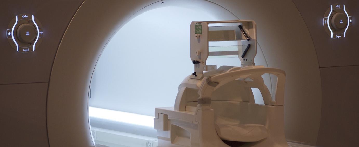
BrainWorks
BrainWorks offers safe and repeatable technologies to researchers for cutting-edge studies of human brain function.
General inquiries