In vivo cancer models
Vish Muthusamy, PhD
About the services
Jump to Cell line-derived xenograft (CDX) models — Patient-derived xenograft (PDX) models — Syngeneic models — Genetically engineered mouse models (GEMMS) — Metastasis models
Cell line-derived xenograft (CDX) models
These experiments typically involve engrafting human cancer cell lines into mice, followed by intervention with a therapeutic agent. Our repository includes a wide range of human cell lines across multiple cancer types, all validated for tumor formation in immunodeficient mouse strains. With comprehensive historic growth data, we can recommend optimal therapeutic windows and define experimental endpoints to demonstrate efficacy. Our experience with standard-of-care drugs in select models also enables the design of combination strategies. Additionally, we can characterize and conduct experiments using investigator-provided engineered cell lines or commercially sourced lines. [Back to top]

Patient-derived xenograft (PDX) models
PDX models are generated using primary human tumor material that has never been exposed to plastic. As a result, these tumors have not undergone the selection pressures associated with in vitro culture and therefore more faithfully recapitulate human tumor biology, excluding the immune compartment. We have extensive experience establishing new PDX models across multiple cancer types and optimizing graft take in immunodeficient host mice. In addition, we have conducted large-scale preclinical efficacy studies of promising therapeutic agents that have subsequently demonstrated clinical success. [Back to top]

Syngeneic models
These models are grafted into fully immunocompetent, syngeneic host mice and are therefore well suited for evaluating therapeutics and strategies that target immune pathways. A substantial portion of PCM’s experience lies in the use of these models, and we can assist in designing rigorous experiments to assess relevant immunotherapies. A wide range of standard-of-care immune-based treatments (checkpoint inhibitors, cytokines, immune cell–specific antibodies, and antibody–drug conjugates) have been tested in these systems, generating data that can be leveraged to develop and evaluate novel combination strategies. PCM is fully equipped to monitor and characterize immune responses throughout the course of these studies, including cytokine profiling and analysis of tumor-infiltrating immune populations, using established platforms and methodologies. [Back to top]


Genetically engineered mouse models (GEMMS)
Genetically engineered mouse models (GEMMs) are designed to recapitulate disease initiation and progression and are powerful tools for understanding tumor biology and disease course. At PCM, we identify the most appropriate GEMM for the scientific question at hand and manage all aspects of model execution, including breeding, genotyping, cohort establishment, and other required manipulations to support rigorous preclinical studies. [Back to top]


Metastasis models
PCM has developed and optimized multiple in vivo models that reflect metastatic disease. These models are particularly valuable for evaluating therapeutic efficacy in advanced-stage cancers involving metastases to distant organs, as well as for assessing potential abscopal effects. Available approaches include grafting or GEMM-based spontaneous metastasis models, vascular introduction of cancer cells and orthotopic implantation. Disease progression and treatment response in these models can be assessed using noninvasive imaging modalities such as bioluminescence, fluorescence, and magnetic resonance imaging or through serial histopathological evaluation. [Back to top]


Available to Yale researchers & external researchers
-
Services
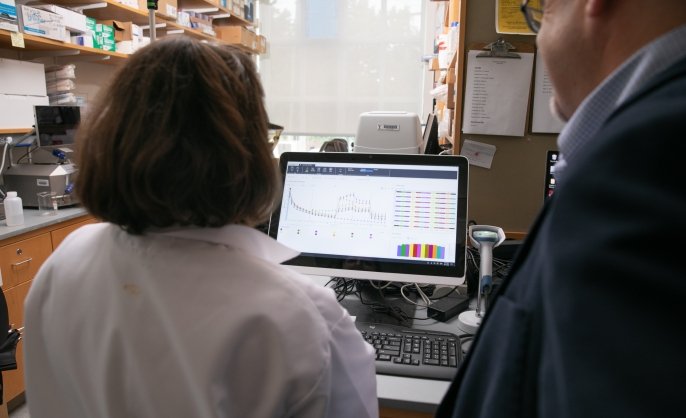
Cellular and mitochondrial respiration and bioenergetics
Cellular and mitochondrial respiration (OCR) and acidification (ECAR) are assessed with the Agilent Seahorse XF Pro 96. In a 96-well plate format, real-time measurements of cellular respiration, oxidative phosphorylation, mitochondrial leak, and mitochondrial transport are assessed as functional kinetic measurements of cellular bioenergetics. Changes in acidification are also observed simultaneously. An integrated Agilent BioTek Cytation5 imager correlates cell number via Hoechst nuclear stain to respiration for more accurate normalization of respiration.
Located within Chemical Metabolism CoreRebecca Cardone, MS
-
Instrument/equipment
BD LSR Fortessa X20
The Fortessa analyzer has 4 lasers, including V 405 nm, B 488 nm, YG 561 nm, and R 640 nm. It can detect 16 colors.
Located within Science Hill Flow Cytometry FacilityFreya Wencker, PhD
-
Instrument/equipment

BD Symphony A1 analyzer
Analyze from single tubes or 96-well plates with 45 lasers (violet, blue, yellow/green, red) and 64 detectors (6 violet, 2 blue, 4 yellow/green, 3 red).
Located within West Campus Flow Cytometry Facility