Cores Directory
Search for the resources you need to answer your research questions.
93 RESULTS FOUND
Listing of Research Core itens
-
Core/Facility
West Campus Analytical Core
Our instruments help characterize and elucidate the structures of chemical and biophysical samples. We train researchers.
-
Core/Facility
West Campus Imaging Core
We offer access to and training in the use of a variety of confocal and epifluorescence microscopes as well as light-sheet, laser microdissection, and atomic force microscopy.
-
Core/Facility
West Campus Materials Characterization Core
We offer a variety of materials characterization tools, including spectroscopy, microscopy, and X-ray fluorescence and diffraction.
-
Instrument/Equipment
Wet benches
The cleanroom houses bench fume hoods that are dedicated to acid use, base use, solvent use, and the RCA cleaning process.
Located within Yale University Cleanroom -
Core/Facility
Yale Institute for Nanoscience and Quantum Engineering (YINQE)
We offer access to electron microscopy, atomic force microscopy, and electron-beam lithography for students, postdocs, and faculty.
Michael Rooks, PhD
Primary contact Associate Director of Facilities Senior Research Scientist in Applied Physics -
Core/Facility
Yale Plant Growth Facility
The core consists of over 20,000 square feet of greenhouses, growth chambers and workspace for plant genetics, evolutionary biology, climate change, and other controlled-environment research.
Christopher Bolick
Primary contact Associate Director of Research, Marsh Botanical Gardens -
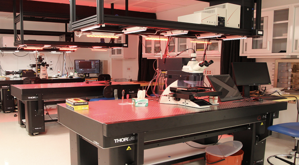
Core/Facility
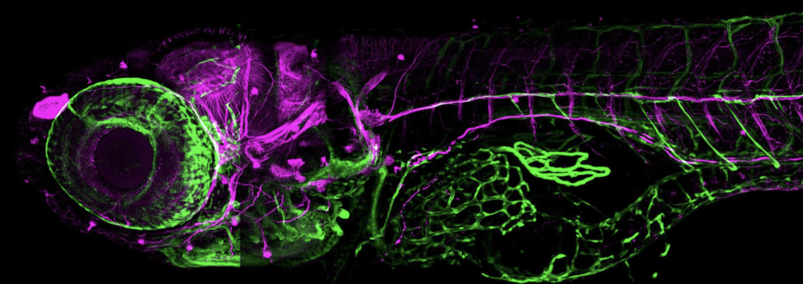
Yale Zebrafish Research Core
The Yale Zebrafish Research Core (YZRC) is a centralized repository of zebrafish services with its own fish facility and experimental space.
-
Instrument/Equipment
Zeiss Lightsheet Z.1 Microscope
A selective plane illumination microscope (SPIM).
Located within Light Microscope Imaging Facility on Science HillJoseph Wolenski, PhD
-
Instrument/Equipment
Zeiss LSM 880 Airyscan Confocal Microscope
Zeiss LSM 880 Airyscan confocal microscope
Located within Confocal Microscopy at CCMI