Cores Directory
Search for the resources you need to answer your research questions.
19 RESULTS FOUND
Active Filters:
Listing of Research Core itens
-
Core/Facility
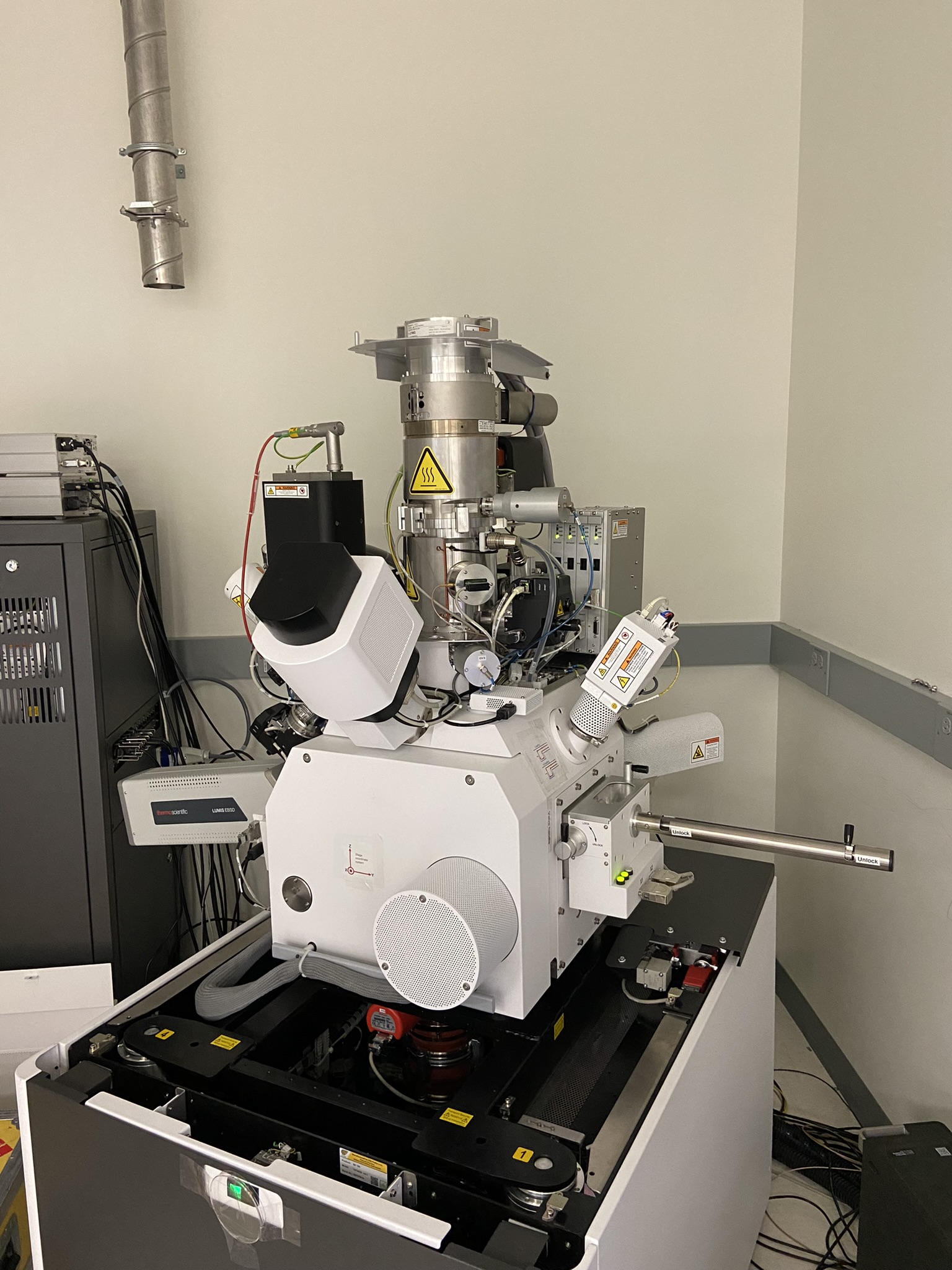
Aberration-Corrected Electron Microscopy (ACEM) Core
Our core's focus is cutting-edge and high-throughput capability in electron microscopy techniques, including TEM, SEM, and FIB.
-
Core/Facility
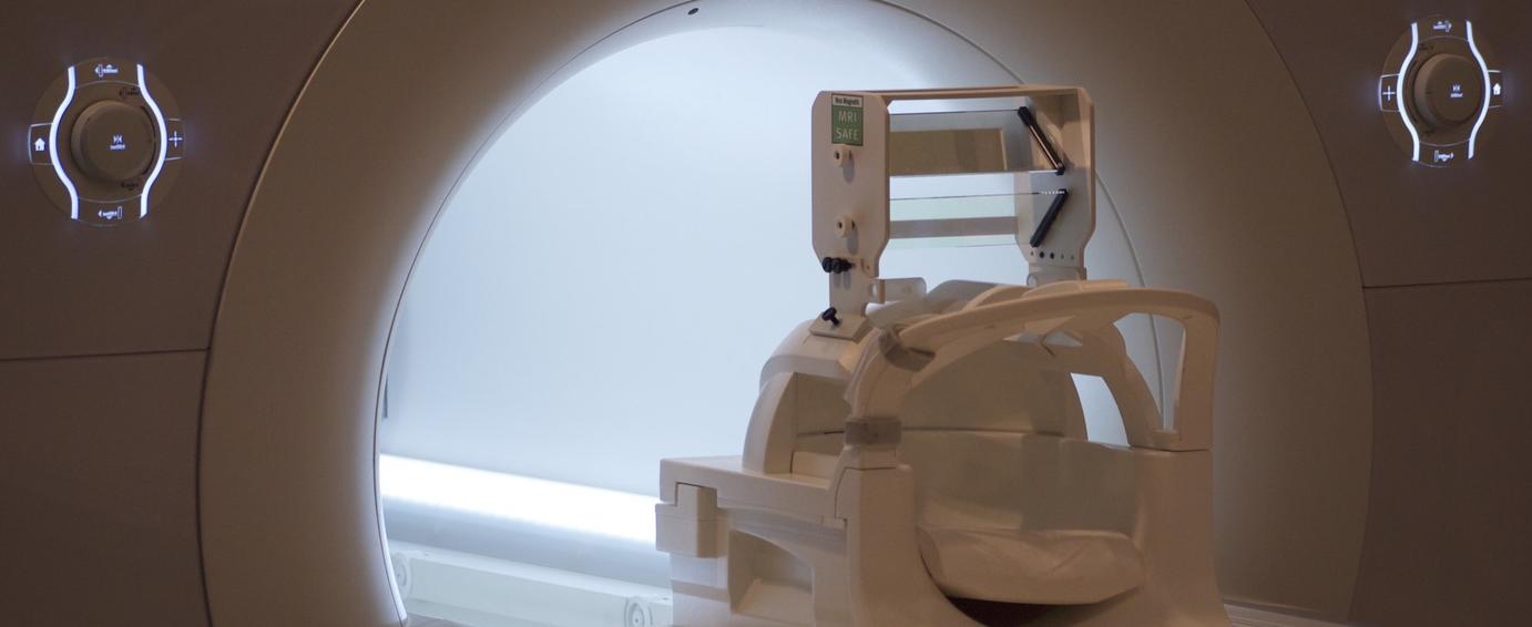
BrainWorks
BrainWorks offers safe and repeatable technologies to researchers for cutting-edge studies of human brain function.
General inquiries
-
Core/Facility
Yale Center for Molecular Discovery
We provide resources for assay development and high-throughput screening with small molecules and arrayed siRNA and sgRNA/CRISPR libraries in biochemical, cell-based and image-based assays (primarily 384-well format).
-
Core/Facility
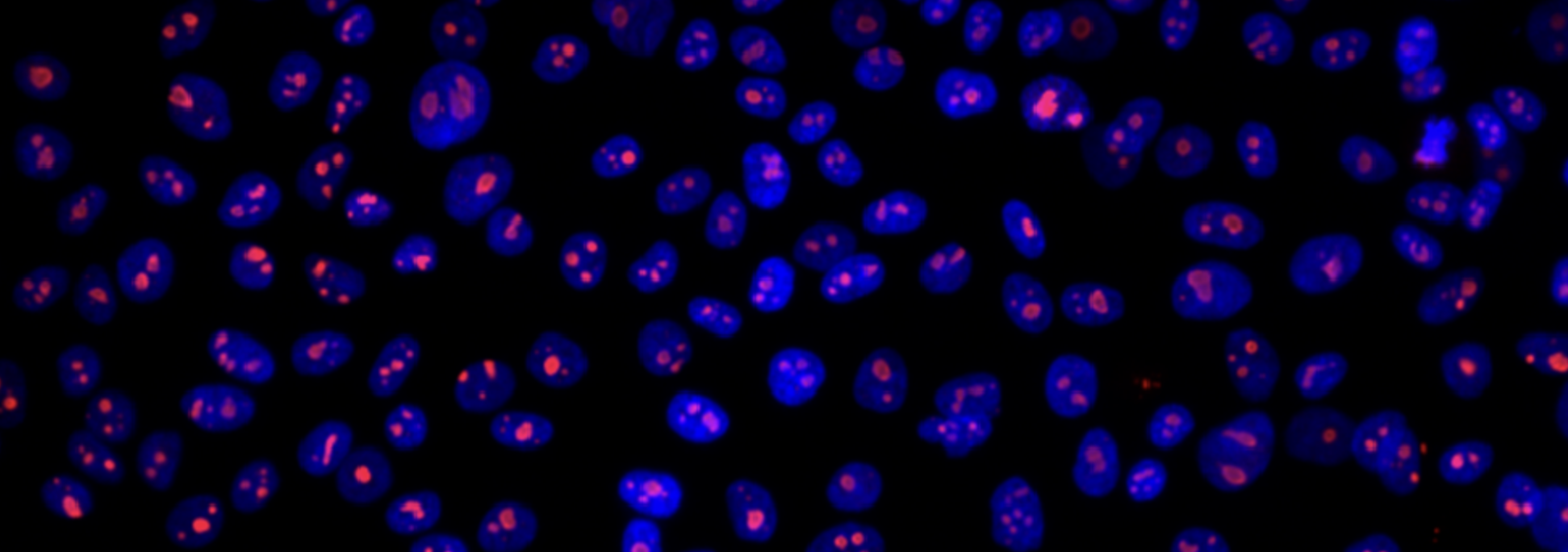
Confocal Microscopy at CCMI
We offer confocal microscopy, two-photon microscopy, light-sheet microscopy, swept-field microscopy, and super-resolution imaging services.
-
Core/Facility
Yale CryoEM Resource
The Yale CryoEM Resource comprises electron microscopes on all 3 campuses: a Titan Krios on West Campus, a Glacios and a Tecnai T12 at Yale School of Medicine, and a Glacios and a Talos L120C on Science Hill.
-
Core/Facility
CyTOF Facility
The CyTOF 2 facility houses a time-of-flight atomic mass spectrometer for the high-speed acquisition of highly multi-parametric single-cell data.
-
Core/Facility
Earth Materials Characterization Center (EMC2)
We offer materials physics equipment including microscopy, spectroscopy, deformation tools, and sample-prep tools for sample characterization work.
-
Core/Facility
Electron Microscopy Facility at CCMI
The Electron Microscopy Facility at the Center for Cellular and Molecular Imaging (CCMI) provides investigators with a wide range of electron microscopy services.
-
Core/Facility
Cores pages FAQ for core directors
Learn to create and edit your core's website on Research at Yale. This page is both a FAQ and a demonstration.
-
Core/Facility
FIB-SEM Collaboration Core (F-SCC)
We utilize unparalleled enhanced FIB-SEM pipeline to generate large-volume three-dimensional electron microscopy data with nanometer isotropic resolution. Such data reveal critical insights about structure-function relationships in biology.
-
Core/Facility
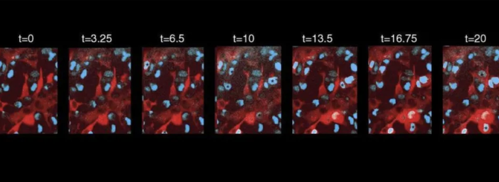
In Vivo Imaging (IVI) Facility
The core offers researchers access to an upright two-photon laser scanning microscope optimized for in vivo fluorescence imaging studies. Training or direct staff assistance is available in surgical preparations, imaging acquisition, and data analysis.
-
Core/Facility
Light Microscope Imaging Facility on Science Hill
We provide training and access to a broad collection of shared confocal microscopes and lab support areas for sample preparation.